. 2010 Jun;31(6):1010-7.
doi: 10.1093/carcin/bgq044. Epub 2010 Feb 22.

Epithelial interleukin-4 receptor expression promotes colon tumor growth

Affiliations

Epithelial interleukin-4 receptor expression promotes colon tumor growth

Felicitas L Koller et al. Carcinogenesis. 2010 Jun.

Abstract

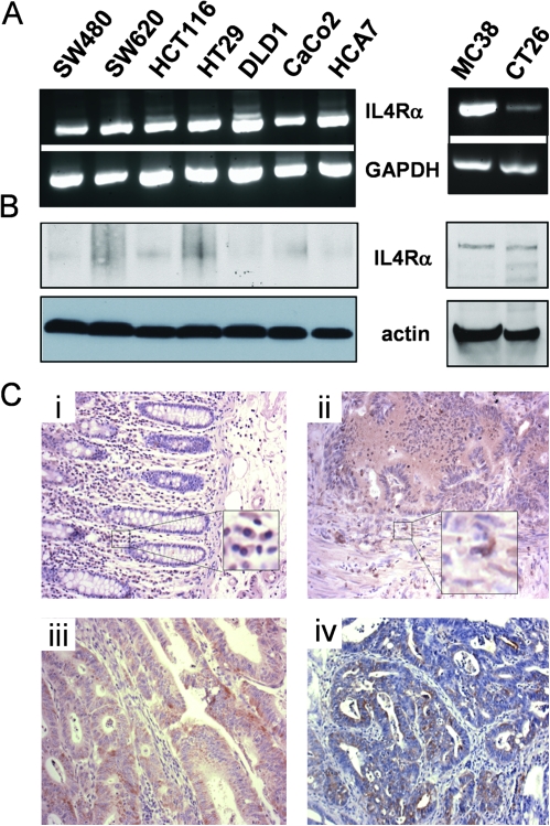
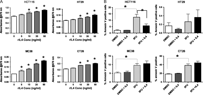
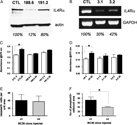
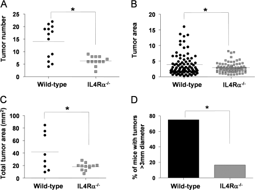
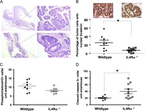
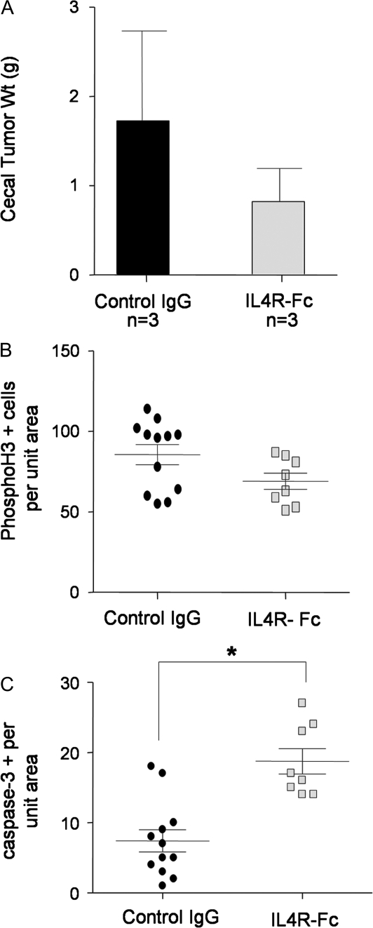
Inflammatory mediators are of considerable interest as potential therapeutic targets in various cancers. Here we investigate whether interleukin (IL)-4 receptor alpha (IL4Ralpha), a component of the receptor complex for the T helper 2 cytokines IL4 and IL13, plays a role in colonic tumorigenesis. IL4Ralpha protein expression was seen in tumor cells of 28/48 human colon adenocarcinomas on a tissue microarray. In human and murine colon tumor cell lines analyzed in vitro, all of which expressed IL4Ralpha, treatment with exogenous ligand resulted in dose-dependent increases in proliferation. IL4 decreased apoptosis only in HCT116 cells. An orthotopic allograft model was used to determine in vivo effects of tumor cell-specific IL4Ra ablation. MC38 murine tumor cells with the IL4Ra gene knocked down showed reduced proliferation but no difference in apoptosis compared with controls after implantation in ceca of syngeneic mice. Mice null for IL4Ra and wild-type controls were treated with azoxymethane and dextran sulfate sodium to induce tumor formation. Mice with global deletion of IL4Ra had significantly fewer and smaller tumors. Reduced tumorigenicity correlated with decreased proliferation and increased apoptosis. Systemic blockade of IL4Ralpha-IL4 interactions with a chimeric soluble receptor protein gave similar results in the cecal implant model. Thus, IL4Ralpha, a component of the IL4R and IL13R, contributes to tumor formation in a mouse model of colitis-associated cancer. Proliferation appears to be directly mediated via IL4Ralpha on the epithelial tumor cells. Survival may be an indirect response mediated via other host cells. Our results support therapeutic targeting of IL4Ralpha in colon cancer.


Figures

Fig. 1.
Fig. 1.
IL4Rα is expressed in human and murine colon cancer cells. (A) IL4Rα (upper panels) and glyceraldehyde3- phosphate dehydrogenase (GAPDH) (lower panels) transcripts in a panel of human (left) and murine (right) colon tumor cell lines as detected by reverse transcription–PCR. (B) IL4Rα (upper panels) and β-actin (lower panels) protein detected by western blotting in lysates from a panel of human (left) and murine (right) cell lines. Note: The antibody for human IL4Rα recognizes only the non-reduced protein; hence, the bands are not as sharp as for the murine protein. (C) Examples of the staining pattern for IL4Rα (brown) seen in tissue sections of (i) normal human colon; (ii, iii) human colon adenocarcinoma on a tissue microarray and (iv) murine AOM-induced colon tumor. Insets show infiltrating cells with positive staining for IL4Rα. Nuclei are counterstained blue with hematoxylin.
Fig. 2.
Fig. 2.
Colon tumor cell lines respond directly to exogenous IL4 in vitro. (A) 3-(4,5-Dimethylthiazole-2-yl)-2,5-biphenyl tetrazolium bromide assays showing the response of four different cell lines, both human (HCT116 and HT29) and mouse (CT26 and MC38), to increasing concentrations of species-specific recombinant IL4. Values shown are for 24 h treatment. Asterisks indicate results significantly higher than the corresponding control (0 ng/ml), P ≤ 0.05 by analysis of variance (ANOVA) with Dunnett's post hoc test. (B) Annexin V assays for cells undergoing apoptosis in response to 24 h exposure to the chemotherapeutic agent, 5-fluorouracil (5FU), in the presence or absence of 20 ng/ml species-specific IL4. Asterisks indicate P ≤ 0.05 by ANOVA with Dunnett's post hoc test.
Fig. 3.
Fig. 3.
IL4Rα knockdown in MC38 and HCT116 cells abrogates proliferative response to IL4. (A) Western blotting of IL4Rα protein (upper panel) and β-actin (lower panel) in MC38 cells infected with control or IL4Ra-targeted shRNA lentivirus. Results are shown for two different targeting sequences. The percentage of expression of IL4Rα, based on actin normalization, is shown below each lane. (B) Reverse transcription–PCR of IL4Rα transcript (upper panel) and GAPDH (lower panel) in HCT116 cells transfected with control or IL4Ra-targeted shRNA plasmids. Results are shown for two different clones. The percentage of expression of IL4Rα, based on GAPDH normalization, is shown below each lane. (C) 3-(4,5-Dimethylthiazole-2-yl)-2,5-biphenyl tetrazolium bromide (MTT) assay of MC38-control and knockdown clones with (black bars) or without (white bars) 24 h treatment with 20 ng/ml murine IL4. Only the control clone showed a significant response. (D) MTT assay of HCT116 control and knockdown clones with or without 20 ng/ml human IL4. (E) Cleaved caspase-3 immunostaining in cecal tumors from mice injected with MC38-control (n = 4) or MC38-knockdown cells (n = 5). Shown are the mean number of positive cells per unit tumor area per mouse, P = 0.95 by Mann–Whitney test. (F) Phosphohistone H3 immunostaining in cecal tumors from mice injected with MC38-control (n = 4) or MC38-knockdown cells (n = 5). Shown are the mean number of positive cells per unit tumor area per mouse, *P = 0.03 by Mann–Whitney test.
Fig. 4.
Fig. 4.
Tumor number and size are significantly reduced in IL4Rα−/− mice following AOM/DSS treatment. (A) Total tumor number per wild-type (n = 13) or IL4Rα−/− (n = 12) colons, *P = 0.0008, Mann–Whitney test. (B) Individual tumor area for wild-type (n = 86 tumors from eight mice) versus IL4Rα−/− (n = 75 tumors from 12 mice), *P = 0.018, Mann–Whitney test. (C) Total tumor area per mouse for wild-type (n = 8) versus IL4Rα−/− (n = 12), *P = 0.015, Mann–Whitney test. (D) Incidence of large tumors (>3 mm diameter) in wild-type (six of eight mice) versus IL4Rα−/− (2 of 12 mice), *P = 0.019, Fisher's exact test.
Fig. 5.
Fig. 5.
Nuclear β-catenin is less prevalent, proliferation is decreased and apoptosis increased in colon tumors from IL4Rα-null mice. (A) Hematoxylin- and eosin-stained tumors from wild-type (top) and ILRα−/− (bottom) mice. Images on the left are taken with a ×2.5 objective, bar = 1 mm. Boxes indicate the region magnified on the right, bar = 100 μm. (B) Quantification of the percentage of tumor cells per mouse examined in which the β-catenin was localized to the nucleus. At least 500 cells were evaluated per mouse, *P = 0.02, Mann–Whitney test. Inset panels show sample immunostaining for β-catenin in tumor cells present in colonic lesions from wild-type (left) and IL4Rα−/− (right) mice. Bar = 65 μm. (C) Phosphohistone H3 immunostaining in colon tumors from wild-type (n = 8) and IL4Rα−/− (n = 8) mice. Shown are the number of positive cells per unit tumor area per mouse, P = 0.06, Mann–Whitney test. (D) Cleaved caspase-3 immunostaining in colon tumors from wild-type (n = 8) and IL4Rα−/− (n = 10) mice. Shown are the number of positive cells per unit tumor area per mouse, *P = 0.02, Mann–Whitney test.
Fig. 6.
Fig. 6.
IL4Rα blockade leads to reduced proliferation and increased apoptosis of tumor cells in an implanted tumor model. (A) Wet weight of MC38 cecal tumors from control immunoglobulin G (IgG) and IL4R-Fc-treated mice. (B) Phosphohistone H3 immunostaining in MC38 cecal tumors from control IgG and IL4R-Fc-treated mice. Shown are the number of positive cells per unit tumor area per section, P = 0.07, Mann–Whitney test. (C) Cleaved caspase-3 immunostaining in MC38 cecal tumors from control IgG and IL4R-Fc-treated mice. Shown are the number of positive cells per unit tumor area per section, *P = 0.003, Mann–Whitney test.

References

    1. Coussens LM, et al. Inflammation and cancer. Nature. 2002;420:860–867. - PMC - PubMed
    1. Mantovani A, et al. Cancer-related inflammation. Nature. 2008;454:436–444. - PubMed
    1. Pohl C, et al. Chronic inflammatory bowel disease and cancer. Hepatogastroenterology. 2000;47:57–70. - PubMed
    1. Itzkowitz SH, et al. Inflammation and cancer IV. Colorectal cancer in inflammatory bowel disease: the role of inflammation. Am. J. Physiol. Gastrointest. Liver Physiol. 2004;287:G7–G17. - PubMed
    1. Grivennikov S, et al. IL-6 and Stat3 are required for survival of intestinal epithelial cells and development of colitis-associated cancer. Cancer Cell. 2009;15:103–113. - PMC - PubMed

Publication types