

Studying the role of human parietal cortex in visuospatial attention with concurrent TMS-fMRI
- PMID: 20176690
- PMCID: PMC2951847
- DOI: 10.1093/cercor/bhq015
Studying the role of human parietal cortex in visuospatial attention with concurrent TMS-fMRI
Abstract
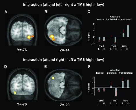
Combining transcranial magnetic stimulation (TMS) with concurrent functional magnetic resonance imaging (fMRI) allows study of how local brain stimulation may causally affect activity in remote brain regions. Here, we applied bursts of high- or low-intensity TMS over right posterior parietal cortex, during a task requiring sustained covert visuospatial attention to either the left or right hemifield, or in a neutral control condition, while recording blood oxygenation-level-dependent signal with a posterior MR surface coil. As expected, the active attention conditions activated components of the well-described "attention network," as compared with the neutral baseline. Also as expected, when comparing left minus right attention, or vice versa, contralateral occipital visual cortex was activated. The critical new finding was that the impact of high- minus low-intensity parietal TMS upon these visual regions depended on the currently attended side. High- minus low-intensity parietal TMS increased the difference between contralateral versus ipsilateral attention in right extrastriate visual cortex. A related albeit less pronounced pattern was found for left extrastriate visual cortex. Our results confirm that right human parietal cortex can exert attention-dependent influences on occipital visual cortex and provide a proof of concept for the use of concurrent TMS-fMRI in studying how remote influences can vary in a purely top-down manner with attentional demands.
Figures


Similar articles
-
Concurrent TMS-fMRI reveals dynamic interhemispheric influences of the right parietal cortex during exogenously cued visuospatial attention.Eur J Neurosci. 2011 Mar;33(5):991-1000. doi: 10.1111/j.1460-9568.2010.07580.x. Epub 2011 Feb 17. Eur J Neurosci. 2011. PMID: 21324004 Free PMC article.
-
Testing the inter-hemispheric competition account of visual extinction with combined TMS/fMRI.Neuropsychologia. 2015 Jul;74:63-73. doi: 10.1016/j.neuropsychologia.2015.04.021. Epub 2015 Apr 21. Neuropsychologia. 2015. PMID: 25911128
-
Interhemispheric effect of parietal TMS on somatosensory response confirmed directly with concurrent TMS-fMRI.J Neurosci. 2008 Dec 3;28(49):13202-8. doi: 10.1523/JNEUROSCI.3043-08.2008. J Neurosci. 2008. PMID: 19052211 Free PMC article.
-
TMS in the parietal cortex: updating representations for attention and action.Neuropsychologia. 2006;44(13):2700-16. doi: 10.1016/j.neuropsychologia.2005.12.007. Epub 2006 Feb 7. Neuropsychologia. 2006. PMID: 16455113 Review.
-
Hemispheric Asymmetry in TMS-Induced Effects on Spatial Attention: A Meta-Analysis.Neuropsychol Rev. 2024 Sep;34(3):838-849. doi: 10.1007/s11065-023-09614-2. Epub 2023 Sep 22. Neuropsychol Rev. 2024. PMID: 37736863 Free PMC article. Review.
Cited by
-
Multisensory stimulation in stroke rehabilitation.Front Hum Neurosci. 2012 Apr 9;6:60. doi: 10.3389/fnhum.2012.00060. eCollection 2012. Front Hum Neurosci. 2012. PMID: 22509159 Free PMC article.
-
Dementia trajectory for patients with logopenic variant primary progressive aphasia.Neurol Sci. 2019 Dec;40(12):2573-2579. doi: 10.1007/s10072-019-04013-z. Epub 2019 Jul 22. Neurol Sci. 2019. PMID: 31332581
-
State-dependent effects of neural stimulation on brain function and cognition.Nat Rev Neurosci. 2022 Aug;23(8):459-475. doi: 10.1038/s41583-022-00598-1. Epub 2022 May 16. Nat Rev Neurosci. 2022. PMID: 35577959 Review.
-
Comparing TMS perturbations to occipital and parietal cortices in concurrent TMS-fMRI studies-Methodological considerations.PLoS One. 2017 Aug 2;12(8):e0181438. doi: 10.1371/journal.pone.0181438. eCollection 2017. PLoS One. 2017. PMID: 28767670 Free PMC article.
-
Causal evidence for frontal involvement in memory target maintenance by posterior brain areas during distracter interference of visual working memory.Proc Natl Acad Sci U S A. 2011 Oct 18;108(42):17510-5. doi: 10.1073/pnas.1106439108. Epub 2011 Oct 10. Proc Natl Acad Sci U S A. 2011. PMID: 21987824 Free PMC article. Clinical Trial.
References
-
- Allen EA, Pasley BN, Duong T, Freeman RD. Transcranial magnetic stimulation elicits coupled neural and hemodynamic consequences. Science. 2007;317:1918–1921. - PubMed
-
- Bestmann S, Ruff CC, Blankenburg F, Weiskopf N, Driver J, Rothwell JC. Mapping causal interregional influences with concurrent TMS-fMRI. Exp Brain Res. 2008;191:383–402. - PubMed
-
- Bestmann S, Ruff CC, Driver J, Blankenburg F. Concurrent TMS and functional magnetic resonance imaging: methods and current advances. In: Wasserman EM, Epstein CM, Ziemann U, Walsh V, Paus T, Lisanby S, editors. The Oxford handbook of transcranial stimulation. Oxford: Oxford University Press; 2008.
-
- Bisley JW, Goldberg ME. Neuronal activity in the lateral intraparietal area and spatial attention. Science. 2003;299:81–86. - PubMed
Publication types
MeSH terms
Grants and funding
LinkOut - more resources
Full Text Sources
Medical
Research Materials
