. 2010 Apr 1;184(7):3554-61.
doi: 10.4049/jimmunol.0903250. Epub 2010 Feb 22.

Depleting syndecan-4+ T lymphocytes using toxin-bearing dendritic cell-associated heparan sulfate proteoglycan-dependent integrin ligand: a new opportunity for treating activated T cell-driven disease

Affiliations

Depleting syndecan-4+ T lymphocytes using toxin-bearing dendritic cell-associated heparan sulfate proteoglycan-dependent integrin ligand: a new opportunity for treating activated T cell-driven disease

Hideo Akiyoshi et al. J Immunol. .

Abstract

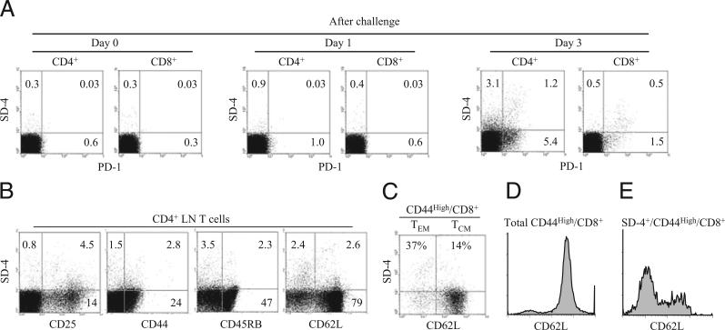
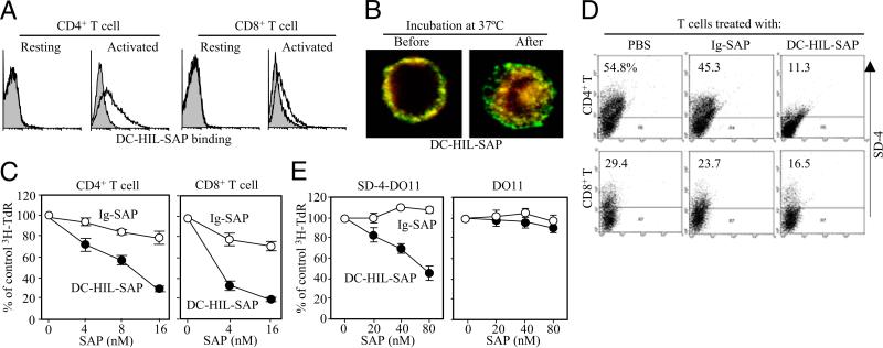
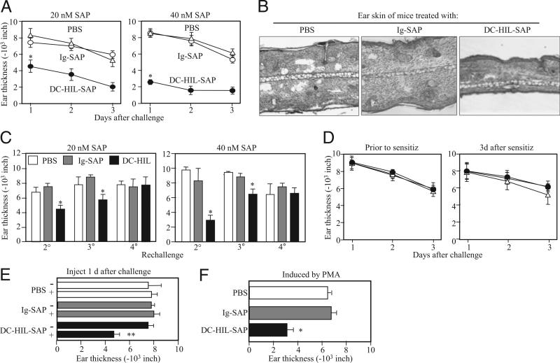
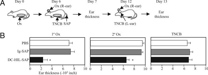
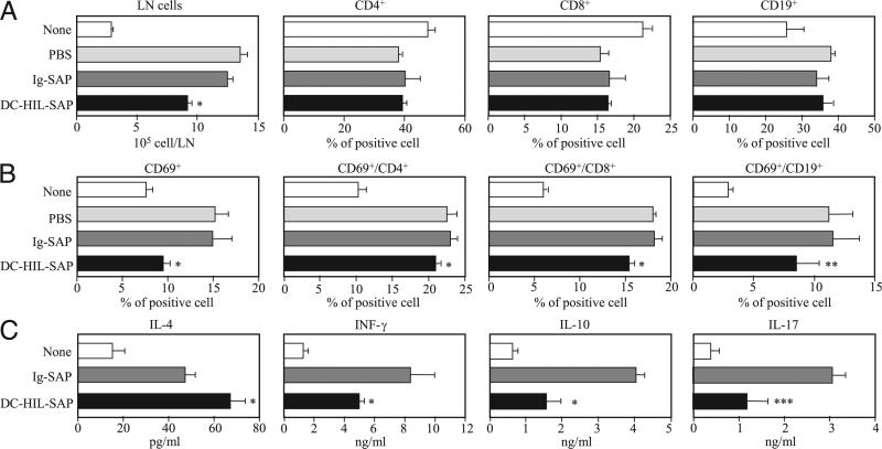
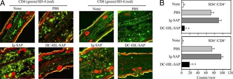
Because syndecan-4 (SD-4) is expressed by some (but not all) T cells following activation and serves as the exclusive ligand of dendritic cell-associated heparan sulfate proteoglycan-dependent integrin ligand (DC-HIL), we envisioned the DC-HIL/SD-4 pathway to be a therapeutic target for conditions mediated by selectively activated T cells. We conjugated soluble DC-HIL receptor with the toxin saporin (SAP; DC-HIL-SAP) and showed it to bind activated (but not resting) T cells and become internalized by and deplete SD-4(+) T cells. In hapten-sensitized mice, DC-HIL-SAP injected i.v. prior to hapten challenge led to markedly suppressed contact hypersensitivity responses that lasted 3 wk and were restricted to the hapten to which the mice were originally sensitized. Such suppression was not observed when DC-HIL-SAP was applied during sensitization. Moreover, the same infusion of DC-HIL-SAP produced almost complete disappearance of SD-4(+) cells in haptenated skin and a 40% reduction of such cells within draining lymph nodes. Our results provide a strong rationale for exploring use of toxin-conjugated DC-HIL to treat activated T cell-driven disease in humans.


Figures

FIGURE 1
FIGURE 1
Characterization of SD-4+ T cells generated in immunized mice. A, CD4+ or CD8+ T cells purified from DLNs of mice sensitized/challenged with Ox day 0 (just prior to challenge), day 1, or day 3 postchallenge were examined for coexpression of SD-4 and PD-1. B, These CD4+ T cells also were analyzed for coexpression of SD-4 and a T cell marker. C–E, Seventeen days postimmunization of OT-I transgenic mice with OVA peptide, CD8+ T cells were purified from DLNs of immunized mice and then stained with FITC–anti-CD44, APC–anti-CD62L, and PE–anti-SD-4 Ab, followed by flow cytometry. C, Dot-blot analysis of SD-4 versus CD62L is shown for CD44high CD8+ cells. CD62Llow and CD62Lhigh fractions represent effector/memory (TEM) and central memory T cells (TCM), respectively. Frequency of SD-4+ cells for each T cell subset is expressed as %. CD62L expression by total CD44high/CD8+ (D) or by SD-4+/CD44high/CD8+ (E) is shown in histograms. All data are representative of at least two experiments.
FIGURE 2
FIGURE 2
Effects of DC-HIL-SAP on T cells in vitro. A, Binding of DC-HIL-SAP to T cells. Splenic CD4+ or CD8+ T cells from BALB/c mice were left untreated (resting) or activated by anti-CD3 Ab (2 μg/ml) for 3 d, and then incubated with 5 μg/ml DC-HIL-SAP (open histograms) or Ig-SAP (shaded), followed by fluorescent labeling with PE–anti-human IgG Ab. Binding was measured by flow cytometry. B, Internalization. Activated CD4+ T cells were incubated with 10 nM DC-HIL-SAP for 1 h on ice. After washing, cells were either left untreated (Before) or incubated (After) at 37°C for 30 min. Cells were then fixed and fluorescently stained with anti-DC-HIL (green fluorescence) or anti-SAP Ab (red), followed by confocal microscopy. Original magnification ×40. C, Effects on T cell proliferation. Splenic CD4+ or CD8+ Tcells(2 × 10⁵ per well) were activated with immobilized anti-CD3 Ab (2 μg/ml) for 2 d, and then different doses of DC-HIL-SAP or Ig-SAP (shown as SAP concentration) were added to the culture. The next day, [3H]thymidine (TdR) incorporation was measured. Relative cpm to the control (0 nM or PBS) are expressed as percentages (mean ± SD; n =3). D, Depletion of SD-4+ T cells. Similarly treated T cells (just prior to harvest for counting [3H] cpm) were examined by flow cytometry for expression of SD-4; percentage of positive cells is shown. E, Specificity to SD-4. DO11.10 T cell lines transfected with SD-4 gene (SD-4-DO11) or empty vector (DO11) were treated with indicated doses of SAP conjugates and cultured for 2 d. Proliferation was measured (mean ± SD; n = 3). Data shown are representative of at least two separate experiments.
FIGURE 3
FIGURE 3
Effects of DC-HIL-SAP on CH. On day 0, BALB/c mice (n = 4) were sensitized by painting 2% Ox on shaved abdominal skin. On day 6, CH was elicited in sensitized mice by painting 1% Ox or solvent control to right or left ears, respectively (Challenge). Ear thickness was measured daily from days 1–3 following challenge. A, Mice also were injected i.v. with PBS, Ig-SAP, or DC-HIL-SAP (20 or 40 nM) 3 h prechallenge. Daily change in ear thickness (10−3 inches) was plotted for each panel. B, On day 2, ear skin specimens of mouse representative in each group were stained with H&E and histologically examined under magnification ×10. C, These mice were kept for 1 wk and then rechallenged with Ox weekly for second (2°), third (3°) and fourth challenges (4°). Ear thickness was measured the day following challenge. D, In separate experiments, mice (n = 4) were also injected i.v. with the same SAP conjugates (40 nM) 3 h presensitization or 3 d postsensitization. E, One day postchallenge, mice (n = 4) were injected with PBS, Ig-SAP, or DC-HIL-SAP (40 nM). Ear thickness was measured just before (−) or 1 d after (+) injection. F, Similarly, mice (n = 4) were also sensitized and challenged but with a different chemical, PMA. Ear thickness was measured 1 d following challenge. Statistical significance is denoted by asterisks (*p < 0.001; **p = 0.003) as compared with ear thickness treated with control Ig-SAP. Data shown are representative of four (A) and two (B–F) independent experiments.
FIGURE 4
FIGURE 4
Suppression of CH by DC-HIL-SAP is restricted to hapten administrated at the time of infusion. A, Immunization protocol is depicted schematically: BALB/c mice (n = 4) were sensitized with Ox on day 0, and i.v. injected with PBS or SAP conjugate 3 h prechallenge (day 6). On the same day, mice were challenged with Ox and solvent alone on right (R-ear) and left ears (L-ear), respectively, and also sensitized to TNCB. On day 7, ear thickness was measured (1° Ox). Day 12, all mice were challenged with Ox (2° Ox) and TNCB on right and left ears, respectively. Ear thickness shown is measured on day 1 every postchallenge (B). *p < 0.05 as compared with ear thickness of mice treated with Ig-SAP. A second experiment showed similar results.
FIGURE 5
FIGURE 5
Immunological phenotypes of DLN cells from mice treated with SAP conjugates. A and B, Sensitized mice (n = 3) were i.v. injected with PBS, Ig-SAP, or DC-HIL-SAP (each 40 nM) 3 h prechallenge. Two days postchallenge with Ox, DLNs procured from treated mice were counted and expressed as cell number per DLN. Frequency (%) of CD4+, CD8+, and CD19+ B cells was measured by flow cytometry (A). DLN cells also were examined for expression of CD69 on all LN cells, CD4+, CD8+, or CD19+ cells, and frequency (%) was calculated (B). C, DLN cells from similarly treated mice (n = 3) were cultured for 2 d in the presence of anti-CD3 and anti-CD28 Ab and assayed for production of IL-4, IFN-γ, IL-10, and IL-17. As control, DLN cells from unsensitized mice (None) were also analyzed in the same manner. SD was derived from experimental values for three mice. Statistical significance is denoted by asterisks (*p < 0.001; ** p = 0.14; *** p = 0.03) as compared with frequency in LNs of mice treated with control Ig-SAP. Data shown are representative of three separate experiments.
FIGURE 6
FIGURE 6
Depletion of SD-4+ T cells in Ox-painted skin. A, BALB/c mice (n = 3) were sensitized, injected with PBS or SAP conjugate, and challenged. Two days postchallenge, ear skin biopsies were procured and doubly stained with anti-CD4 or CD8 Ab (shown in green fluorescence) and anti–SD-4 Ab (red). Original magnification ×10. Merged confocal images are shown. B, Using these images, numbers of SD-4+/CD4+ or SD-4+/CD8+ T cells were counted in three separate views, and the average with SD is shown graphically. *p < 0.01 as compared with percentage in skin of mice infused with Ig-SAP. Data shown are representative of three separate experiments.
FIGURE 7
FIGURE 7
DC-HIL-SAP deletes SD-4+ T cells within DLNs. Mice (n = 3) were sensitized, injected, and challenged similarly. Two days post-challenge, DLN cells were isolated from treated mice and pooled, from which CD4+ or CD8+ T cells were purified and examined for expression of SD-4 by flow cytometry. A, The frequency (%) is shown in dot blots. B, Total number of SD-4+/CD4+ or SD-4+/CD8+ T cells per DLN was calculated and shown graphically. A second experiment showed similar results.

Similar articles

Cited by

References

    1. Chambers CA, Allison JP. Co-stimulation in T cell responses. Curr. Opin. Immunol. 1997;9:396–404. - PubMed
    1. Carreno BM, Collins M. The B7 family of ligands and its receptors: new pathways for costimulation and inhibition of immune responses. Annu. Rev. Immunol. 2002;20:29–53. - PubMed
    1. Carreno BM, Bennett F, Chau TA, Ling V, Luxenberg D, Jussif J, Baroja ML, Madrenas J. CTLA-4 (CD152) can inhibit T cell activation by two different mechanisms depending on its level of cell surface expression. J. Immunol. 2000;165:1352–1356. - PubMed
    1. Linsley PS, Brady W, Urnes M, Grosmaire LS, Damle NK, Ledbetter JA. CTLA-4 is a second receptor for the B cell activation antigen B7. J. Exp. Med. 1991;174:561–569. - PMC - PubMed
    1. Tivol EA, Borriello F, Schweitzer AN, Lynch WP, Bluestone JA, Sharpe AH. Loss of CTLA-4 leads to massive lymphoproliferation and fatal multiorgan tissue destruction, revealing a critical negative regulatory role of CTLA-4. Immunity. 1995;3:541–547. - PubMed

Publication types

MeSH terms