Review
. 2010 May;78(5):1809-23.
doi: 10.1128/IAI.01384-09. Epub 2010 Feb 22.

Phylogenomics reveals a diverse Rickettsiales type IV secretion system

Affiliations
Review

Phylogenomics reveals a diverse Rickettsiales type IV secretion system

Joseph J Gillespie et al. Infect Immun. 2010 May.

Abstract

With an obligate intracellular lifestyle, Alphaproteobacteria of the order Rickettsiales have inextricably coevolved with their various eukaryotic hosts, resulting in small, reductive genomes and strict dependency on host resources. Unsurprisingly, large portions of Rickettsiales genomes encode proteins involved in transport and secretion. One particular transporter that has garnered recent attention from researchers is the type IV secretion system (T4SS). Homologous to the well-studied archetypal vir T4SS of Agrobacterium tumefaciens, the Rickettsiales vir homolog (rvh) T4SS is characterized primarily by duplication of several of its genes and scattered genomic distribution of all components in several conserved islets. Phylogeny estimation suggests a single event of ancestral acquirement of the rvh T4SS, likely from a nonalphaproteobacterial origin. Bioinformatics analysis of over 30 Rickettsiales genome sequences illustrates a conserved core rvh scaffold (lacking only a virB5 homolog), with lineage-specific diversification of several components (rvhB1, rvhB2, and rvhB9b), likely a result of modifications to cell envelope structure. This coevolution of the rvh T4SS and cell envelope morphology is probably driven by adaptations to various host cells, identifying the transporter as an important target for vaccine development. Despite the genetic intractability of Rickettsiales, recent advancements have been made in the characterization of several components of the rvh T4SS, as well as its putative regulators and substrates. While current data favor a role in effector translocation, functions in DNA uptake and release and/or conjugation cannot at present be ruled out, especially considering that a mechanism for plasmid transfer in Rickettsia spp. has yet to be proposed.


Figures

FIG. 1.
FIG. 1.
P-T4SSs. (A) Model of the vir P-T4SS encoded on the pTi plasmid of A. tumefaciens. B1 to B11, VirB1 to VirB11; D4, VirD4. (B) Comparison of the rvh P-T4SSs from Rickettsiales with similar P-T4SSs from other bacteria. At vir, A. tumefaciens Ti plasmid P-T4SS; Ec tra, E. coli IncN plasmid pKM101 P-T4SS; Ec trw, E. coli plasmid R388 P-T4SS; Lp lvh, L. pneumophila P-T4SS; Bs vir, Brucella suis P-T4SS; and Bp ptl, B. pertussis P-T4SS. VirB1 of B. pertussis is depicted with the N-terminal glycohydrolase domain of PtlE (ntd-E) (107). The rvh examples are shown within the dashed-line box: Rt rvh, R. typhi P-T4SS; Ot rvh, O. tsutsugamushi P-T4SS; Ns rvh, Neorickettsia sennetsu P-T4SS; Wp rvh, Wolbachia pipientis P-T4SS; Ap rvh, A. phagocytophilum P-T4SS; and Er rvh, Ehrlichia ruminantium P-T4SS. X indicates that no gene for the component has been annotated and no subjects were detectable using tblastn; P represents the proliferation of rvhB2 genes, putative VirB2-like encoding genes.
FIG. 2.
FIG. 2.
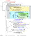
Estimated phylogeny for 47 diverse P-T4SSs. Taxa are colored according to proteobacterial class: red, Epsilonproteobacteria; blue, Gammaproteobacteria; black, Alphaproteobacteria; and green, Betaproteobacteria. Each taxon name is appended with its P-T4SS nomenclature and a number (I, II, III, or IV) referring to recently proposed categories of P-T4SSs (93). Plasmid-encoded P-T4SSs are indicated by yellow circles. The 18 sampled Rickettsiales taxa are within a gray box. Rickettsia spp. are shown as a cladogram due to limited sequence divergence relative to that in the remaining taxa, with the following abbreviations: AG, ancestral group; TG, typhus group; TRG, transitional group; and SFG, spotted fever group (56, 57). The tree is from two independent Bayesian analyses of six P-T4SS proteins (VirB4, VirB8 to VirB11, and VirD4). The topology of the sampled tree with the greatest likelihood (Lnl = −201,623.248) is shown, with branch support assessed from probabilities of clade occurrence in the posterior distribution of 1,906 sampled trees. Asterisks indicate probabilities of 100%. For Wolbachia species, the species in which they are symbionts are listed in parentheses (D. ananassae, Drosophila ananassae; D. melanogaster, Drosophila melanogaster). C. j. jejuni, C. jejuni subsp. jejuni; T. adhaerens, “Trichoplax adhaerens”; L. p. pneumophila, L. pneumophila subsp. pneumophila; X. fastidiosa, Xylella fastidiosa; A. punctata, Aeromonas punctata; X. c. campestris, Xanthomonas campestris pv. campestris; C. coli, Campylobacter coli; Y. pestis, Yersinia pestis; S. meliloti, Sinorhizobium meliloti; V. fischeri, Vibrio fischeri; S. typhimurium, Salmonella enterica serovar Typhimurium; E. amylovora, Erwinia amylovora; P. s. syringae, Pseudomonas syringae subsp. syringae; B. henselae, Bartonella henselae.
FIG. 3.
FIG. 3.
Diversification in rvh T4SS architecture across the major lineages of Rickettsiales. The schematic at the top depicts the deletion and duplication events that possibly occurred prior to the split of the major Rickettsiales lineages (corresponding to the yellow box at the root of the cladogram). The color scheme for the rvh components is consistent throughout the figure and reflects the five islets (A to E) of the Rickettsia rvh P-T4SS described previously (55). The cladogram is simplified from data obtained by phylogeny estimation across 31 rvh P-T4SSs (see Fig. S1 in the supplemental material). Colored boxes in the cladogram depict deviations from the ancestral rvh P-T4SS and are explained at the bottom. Synteny maps (shaded inset) are shown for select taxa. Note that in Rickettsiales other than Rickettsia spp., rvhB9a, rvhB8a, and rvhB7 are removed from islet C. Also, multiple rvhB2 orthologs per genome are depicted, with corresponding numbers in parentheses.
FIG. 4.
FIG. 4.
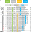
P-T4SS lipoproteins. (A) Schematic depiction of typical characteristics of VirB7 and related proteins. The color scheme is used to identify distinct features in the alignment (see the text for explanation). (B) Manual alignment of 13 diverse VirB7 and VirB7-like lipoproteins with 23 RvhB7 lipoproteins (the latter are boxed). Coordinates for each sequence are shown to the right, with numbers in parentheses to the left referring to flanking residues not shown in the alignment. Protein annotation is shown to the left. The black horizontal bar over the sequences of the predicted processed lipoproteins identifies the region of the NMR structure for the interaction of the TraO C-terminal domain (VirB9-like protein) and TraN (VirB7), both encoded by plasmid pKM101 in E. coli. Sequences above the orange line depict lipoproteins without a predicted Cys-Cys interaction with VirB9 and VirB9-like proteins: Ec, E. coli TraN, encoded by plasmid pKM101 (accession no. NP_511194); Ec2, E. coli TrwH protein (accession no. FAA00034); Bh, B. henselae TrwH-like protein (accession no. AAM82208); Bp, B. pertussis TraI protein, encoded by plasmid pSB102 (accession no. NP_361043); Aa, Aggregatibacter actinomycetemcomitans lipoprotein (accession no. NP_067577); Bp2, B. pertussis putative bacterial secretion system protein (accession no. NP_882291); Ps, P. syringae subsp. syringae VirB7 (accession no. NP_940729); and Bs, B. suis VirB7 (accession no. AAN33275). Sequences below the orange line represent proteins predicted to interact with VirB9 and VirB9-like proteins via a Cys-Cys bond: At, A. tumefaciens VirB7 (accession no. NP_536291); At2, A. tumefaciens AvhB7 (accession no. NP_396098); Xc, X. citri VirB7 (accession no. NP_942612); Br, R. bellii strain RML 369-C RvhB7 (accession no. YP_538183); Bo, R. bellii strain OSU 85 389 RvhB7 (accession no. YP_001495873); Ca, R. canadensis strain McKiel RvhB7 (accession no. YP_001492545); Pr, R. prowazekii strain Madrid E RvhB7 (accession no. NP_220672); Ty, R. typhi strain Wilmington RvhB7 (accession no. YP_067241); Fe, R. felis strain URRWXCal2 RvhB7 (accession no. YP_246480); Ak, R. akari strain Hartford RvhB7 (accession no. YP_001493229); Ma, R. massiliae strain MTU5 RvhB7 (accession no. YP_001499186); Ri, R. rickettsii strain Sheila Smith RvhB7 (accession no. YP_001494506); Rw, R. rickettsii strain Iowa RvhB7 (accession no. YP_001649753); Co, R. conorii strain Malish 7 RvhB7 (accession no. NP_360023); Si, R. sibirica strain 246 RvhB7 (accession no. ZP_00142155); Af, R. africae strain ESF-5 RvhB7 (accession no. ZP_02336216); Pp, protein of the Wolbachia symbiont of C. quinquefasciatus (accession no. YP_001975581); Me, protein of the Wolbachia symbiont of D. melanogaster; Bm, protein of the Wolbachia symbiont of Brugia malayi; Mg, A. marginale protein (accession no. YP_153652); Ph, A. phagocytophilum protein; Rg, E. ruminantium strain Gardel protein; We, E. ruminantium strain Welgevonden (Erwe) protein; Wo, E. ruminantium strain Welgevonden (Erum) protein; Ck, E. chaffeensis strain Arkansas protein; Cj, E. canis strain Jake protein; Hp, H. pylori protein (accession no. CAA10654); and Cje, C. jejuni protein (accession no. NP_863349). RvhB7 taxon codes colored red indicate that sequences corresponding to putative unannotated ORFs were recovered from tblastn searches of the Anaplasmataceae database (sequence coordinates are listed in Table S2 in the supplemental material). Additional conserved Cys residues in the RvhB7 sequences are in bold. A second putative PhN+ motif in the Rickettsia RvhB7 sequences is shaded in blue.
FIG. 5.
FIG. 5.
P-T4SS structural and functional diversification. The dashed-line arrows illustrate the mode of secretion, with substrates depicted by an encircled “s.” The color scheme of P-T4SS components VirB1 (green), VirB2 (light orange), VirB5 (dark orange), VirB7 (yellow), VirB9 (blue), VirB10 (dark blue), and VirD4 (pink) across four secretion systems implies homology. Distinct N and C termini of VirB10 and VirD4 are depicted. M, murein layer. (A) Model of transport for the A. tumefaciens vir P-T4SS. The two-step process of substrate attachment (left) and substrate transfer upon sloughing off of the T-pilus (right) is shown (51). (B) Model of transport for the B. pertussis ptl P-T4SS. VirB1 is distinguished to depict the N-terminal glycohydrolase domain of PtlE, a VirB8 homolog (107). (C) Model of transport for the Rickettsia rvh P-T4SS. (D) General model of transport for the O. tsutsugamushi rvh P-T4SS and for P-T4SSs in species of Anaplasmataceae that are not predicted to completely synthesize PG and LPS.

Similar articles

Cited by

References

    1. Alvarez-Martinez, C. E., and P. J. Christie. 2009. Biological diversity of prokaryotic type IV secretion systems. Microbiol. Mol. Biol. Rev. 73:775-808. - PMC - PubMed
    1. Aly, K. A., and C. Baron. 2007. The VirB5 protein localizes to the T-pilus tips in Agrobacterium tumefaciens. Microbiology 153:3766-3775. - PubMed
    1. Amano, K., A. Tamura, N. Ohashi, H. Urakami, S. Kaya, and K. Fukushi. 1987. Deficiency of peptidoglycan and lipopolysaccharide components in Rickettsia tsutsugamushi. Infect. Immun. 55:2290-2292. - PMC - PubMed
    1. Anderson, L. B., A. V. Hertzel, and A. Das. 1996. Agrobacterium tumefaciens VirB7 and VirB9 form a disulfide-linked protein complex. Proc. Natl. Acad. Sci. U. S. A. 93:8889-8894. - PMC - PubMed
    1. Andersson, S. G., A. Zomorodipour, J. O. Andersson, T. Sicheritz-Ponten, U. C. Alsmark, R. M. Podowski, A. K. Naslund, A. S. Eriksson, H. H. Winkler, and C. G. Kurland. 1998. The genome sequence of Rickettsia prowazekii and the origin of mitochondria. Nature 396:133-140. - PubMed

Publication types

LinkOut - more resources