. 2010 May;78(5):1895-904.
doi: 10.1128/IAI.01165-09. Epub 2010 Feb 22.

The Rickettsia conorii autotransporter protein Sca1 promotes adherence to nonphagocytic mammalian cells

Affiliations

The Rickettsia conorii autotransporter protein Sca1 promotes adherence to nonphagocytic mammalian cells

Sean P Riley et al. Infect Immun. 2010 May.

Abstract

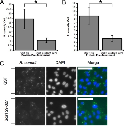
The pathogenesis of spotted fever group (SFG) Rickettsia species, including R. conorii and R. rickettsii, is acutely dependent on adherence to and invasion of host cells, including cells of the mammalian endothelial system. Bioinformatic analyses of several rickettsia genomes revealed the presence of a cohort of genes designated sca genes that are predicted to encode proteins with homology to autotransporter proteins of Gram-negative bacteria. Previous work demonstrated that three members of this family, rOmpA (Sca0), Sca2, and rOmpB (Sca5) are involved in the interaction with mammalian cells; however, very little was known about the function of other conserved rickettsial Sca proteins. Here we demonstrate that sca1, a gene present in nearly all SFG rickettsia genomes, is actively transcribed and expressed in R. conorii cells. Alignment of Sca1 sequences from geographically diverse SFG Rickettsia species showed that there are high degrees of sequence identity and conservation of these sequences, suggesting that Sca1 may have a conserved function. Using a heterologous expression system, we demonstrated that production of R. conorii Sca1 in the Escherichia coli outer membrane is sufficient to mediate attachment to but not invasion of a panel of cultured mammalian epithelial and endothelial cells. Furthermore, preincubation of a recombinant Sca1 peptide with host cells blocked R. conorii cell association. Together, these results demonstrate that attachment to mammalian cells can be uncoupled from the entry process and that Sca1 is involved in the adherence of R. conorii to host cells.


Figures

FIG. 1.
FIG. 1.
Sca1 is expressed in cultured R. conorii Malish7. (A) RT-PCR detection of sca1 mRNA transcript from R. conorii cultured in Vero cells. Specific sca1 cDNA was amplified only after incubation with both input RNA and reverse transcriptase (RTase) in order to eliminate the possibility of exogenous DNA contamination in the reverse transcription reaction. As a positive control we amplified ompA, a gene known to be expressed under culture conditions. (B) Total lysate from purified R. conorii was separated by SDS-PAGE. Protein species that migrated slower than the readily visible rOmpB protein (120 kDa) were excised from the gel and subjected to microcapillary LC-MS/MS peptide sequencing. For reference, a higher magnification of the sequenced portion of the SDS-PAGE gel is shown to the right of the entire Coomassie blue-stained gel. The recovered peptide sequence hits encompassed many of the predicted high-molecular-weight proteins from R. conorii, including rOmpA, rOmpB, and Sca1 (see Table S1 in the supplemental material). Sca1 was the most abundant protein sequenced in this sample as determined by the number of peptides sequenced and the extent of protein coverage. (C) Western immunoblot analysis of total R. conorii protein lysates using rabbit anti-Sca1(29-327) serum (α-Sca1) detected a protein with a molecular mass of approximately 130 kDa (lane 1) that corresponds to the putatively processed Sca1 protein but is not reactive with a total protein lysate from uninfected Vero cells (lane 2). Normal rabbit serum (NRS) did not detect Sca1. (D) Specificity of anti-Sca1 antisera. Recombinant GST-Sca1(29-327) (lane 1) and the 10-histidine-tagged rOmpB passenger domain (amino acids 35 to 1334) (lane 2) were separated by SDS-PAGE, stained with Coomassie blue (left panel), and immunoblotted with the antisera indicated [anti-Sca1(29-327) serum [α-Sca1] and normal rabbit serum (NRS)]. Sca1 antiserum detects recombinant Sca1 but not rOmpB. (E) Flow cytometric analysis of R. conorii. Purified R. conorii was stained with murine anti-R. conorii antiserum and anti-mouse IgG PE-conjugated antisera. Cells were then counterstained with either normal rabbit serum or rabbit anti-Sca1 (26-327) and FITC-conjugated anti-rabbit IgG. PE-positive cells were gated and then analyzed for FITC expression. Cells incubated with normal rabbit serum produced only background levels of fluorescence (red trace), while for cells incubated with anti-Sca1 antisera there was a significant increase in fluorescence intensity indicative of Sca1 surface expression (blue trace). (F) Immunofluorescence microscopy using specific anti-Sca1(29-327) revealed the presence of Sca1. The panels on the left show mouse anti-R. conorii hyperimmune serum recognition of whole bacteria. The panels in the middle show recognition by samples of rabbit serum, including anti-Sca1(29-327) and normal rabbit serum (NRS), and the panels on the right show merged images of appropriate rabbit and mouse serum signals. Sca1 appears to be present in distinct foci on the R. conorii outer membrane.
FIG. 2.
FIG. 2.
Expression of R. conorii Sca1 on the surface of E. coli. (A) Diagram of the sca1-containing pET-22b plasmid variant pSca1-200, showing the relevant 5′ and 3′ features. This vector encodes a recombinant protein fusion containing an N-terminal E. coli PelB signal sequence, R. conorii Sca1, and a C-terminal His6 tag. Due to the N-terminal PelB signal sequence and the inherent ability of autotransporter proteins to direct their own translocation across the bacterial outer membrane, Sca1 is predicted to be anchored in the bacterial outer membrane and primarily exposed to the extracellular environment. RBS, ribosome binding site. (B) Western blot analysis of outer membrane preparations of E. coli harboring the empty vector pET22b, pSca1-200, or ompB-containing pYC9 with anti-Sca1 (amino acids 144 to 157) or polyclonal anti-rOmpB. The presence of pYC9 has previously been demonstrated to permit rOmpB expression. Antibodies against Sca1 and rOmpB are not cross-reactive. “T7 pro” and “T7 term” refer to the transcriptional promoter and terminator sequences, respectively, present in pET-22b.
FIG. 3.
FIG. 3.
Sca1-expressing E. coli adheres to mammalian cells. (A) E. coli BL21(DE3)(pSca1-200) cells were induced using distilled water (top row) or 0.5 mM IPTG (bottom row). These bacteria were incubated with Vero cells, and the preparations were washed to remove nonadherent bacteria. The cells were then permeabilized and stained with the antibodies or fluorophores indicated. The results are typical of three separate experiments. Scale bars = 100 μm. αE. coli, anti-E. coli. (B, C, and D) In lieu of immunofluorescent staining, E. coli cells harboring pET22b (empty vector) or pSca1-200 and induced with IPTG were enumerated by plate-based quantification of CFU that adhered to HeLa (B), Vero (C) or EAhy.926 (D) cells. All results are expressed as percentages of adherent bacteria based on the total bacterial input. The data are representative of the data from at least 3 independent assays for each host cell line. *, P ≤ 0.01.
FIG. 4.
FIG. 4.
Sca1-expressing E. coli cells do not invade mammalian cells. Sca1-expressing E. coli cells were incubated with the HeLa (A), Vero (B), or EAhy.926 (C) mammalian cell lines, washed with PBS to remove nonadherent bacteria, and then treated with gentamicin to kill extracellular bacteria. The remaining bacteria were enumerated. All experiments were conducted with rOmpB-expressing E. coli(pYC9) as a positive control for invasion of mammalian cells. The results are expressed as percentages of the input CFU and are typical of the results of three separate experiments.
FIG. 5.
FIG. 5.
Incubation with a recombinant GST-Sca1 peptide blocks interaction with mammalian cells. Preincubation of Vero (A and C) or EAhy.926 (B) cells with rGST-Sca1(29-327) but not preincubation with rGST-His blocks association of R. conorii with host cells. After incubation of the peptides with the host cells, R. conorii cells were added to the mixtures, and contact was induced by centrifugation. After incubation, the cells were extensively washed and stained for immunofluorescent analysis with anti-R. conorii and DAPI to visualize the host nuclei (C). The remaining R. conorii cells were enumerated by immunofluorescence, and the results are expressed as the ratios of R. conorii cells to host cells (nuclei) (A and B). The data are representative of the data from at least three independent experiments. *, P < 0.01. Scale bars = 50 mm.
FIG. 6.
FIG. 6.
Model for the initial interactions of R. conorii with host cells. SFG Rickettsia species must enter host cells in order to survive. The two initial functions that must be performed by the bacteria are adherence to and invasion of the host cells. Adherence is mediated in part by the rickettsial proteins Sca1, Sca2, rOmpA, and rOmpB and possibly by other factors, such as other conserved Sca proteins. In contrast, Sca2 and rOmpB have been demonstrated to promote internalization. rOmpB is sufficient to trigger internalization in the absence of other virulence factors using pathways involving actin polymerization, clathrin, caveolin, and Ku70. The adherence and invasion processes are therefore mediated by many rickettsial factors, and disruption of any single receptor-ligand interaction is unlikely to completely abrogate infection by SFG Rickettsia species. Furthermore, since multiple proteins promote the same functions, we cannot eliminate the possibility that larger heteromeric protein complexes are involved in the adherence and invasion processes.

Similar articles

Cited by

References

    1. Billings, A. N., H. M. Feng, J. P. Olano, and D. H. Walker. 2001. Rickettsial infection in murine models activates an early anti-rickettsial effect mediated by NK cells and associated with production of gamma interferon. Am. J. Trop. Med. Hyg. 65:52-56. - PubMed
    1. Blanc, G., M. Ngwamidiba, H. Ogata, P. E. Fournier, J. M. Claverie, and D. Raoult. 2005. Molecular evolution of rickettsia surface antigens: evidence of positive selection. Mol. Biol. Evol. 22:2073-2083. - PubMed
    1. Braun, L., H. Ohayon, and P. Cossart. 1998. The InIB protein of Listeria monocytogenes is sufficient to promote entry into mammalian cells. Mol. Microbiol. 27:1077-1087. - PubMed
    1. Cardwell, M. M., and J. J. Martinez. 2009. The Sca2 autotransporter protein from Rickettsia conorii is sufficient to mediate adherence to and invasion of cultured mammalian cells. Infect. Immun. 77:5272-5280. - PMC - PubMed
    1. Chan, Y. G., M. M. Cardwell, T. M. Hermanas, T. Uchiyama, and J. J. Martinez. 2009. Rickettsial outer-membrane protein B (rOmpB) mediates bacterial invasion through Ku70 in an actin, c-Cbl, clathrin and caveolin 2-dependent manner. Cell. Microbiol. 11:629-644. - PMC - PubMed

Publication types

MeSH terms

Substances