. 2010 Mar 15;207(3):535-52.
doi: 10.1084/jem.20092121. Epub 2010 Feb 22.

Bleomycin and IL-1beta-mediated pulmonary fibrosis is IL-17A dependent

Affiliations

Bleomycin and IL-1beta-mediated pulmonary fibrosis is IL-17A dependent

Mark S Wilson et al. J Exp Med. .

Abstract

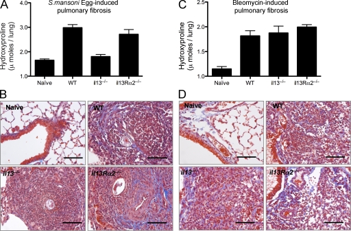
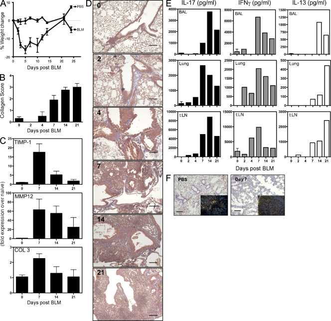
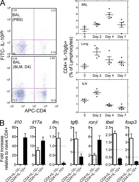
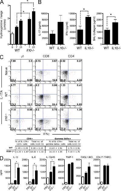
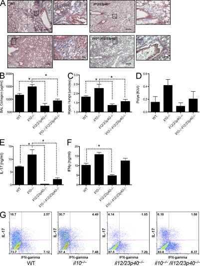
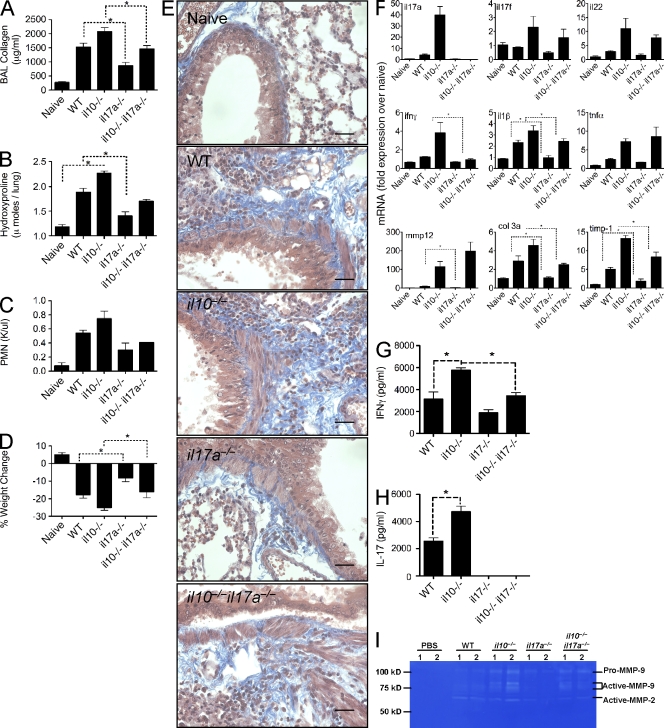
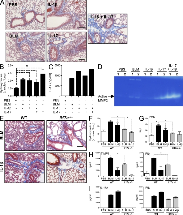
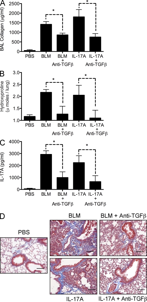
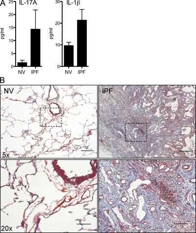
Idiopathic pulmonary fibrosis (IPF) is a destructive inflammatory disease with limited therapeutic options. To better understand the inflammatory responses that precede and concur with collagen deposition, we used three models of pulmonary fibrosis and identify a critical mechanistic role for IL-17A. After exposure to bleomycin (BLM), but not Schistosoma mansoni eggs, IL-17A produced by CD4(+) and gammadelta(+) T cells induced significant neutrophilia and pulmonary fibrosis. Studies conducted with C57BL/6 il17a(-/-) mice confirmed an essential role for IL-17A. Mechanistically, using ifngamma(-/-), il10(-/-), il10(-/-)il12p40(-/-), and il10(-/-)il17a(-/-) mice and TGF-beta blockade, we demonstrate that IL-17A-driven fibrosis is suppressed by IL-10 and facilitated by IFN-gamma and IL-12/23p40. BLM-induced IL-17A production was also TGF-beta dependent, and recombinant IL-17A-mediated fibrosis required TGF-beta, suggesting cooperative roles for IL-17A and TGF-beta in the development of fibrosis. Finally, we show that fibrosis induced by IL-1beta, which mimics BLM-induced fibrosis, is also highly dependent on IL-17A. IL-17A and IL-1beta were also increased in the bronchoalveolar lavage fluid of patients with IPF. Together, these studies identify a critical role for IL-17A in fibrosis, illustrating the potential utility of targeting IL-17A in the treatment of drug and inflammation-induced fibrosis.


Figures

Figure 1.
Figure 1.
IL-13–dependent and –independent pulmonary fibrosis. WT, il13−/−, or il13Rα2−/− mice were given either 5,000 S. mansoni eggs i.p. followed by 5,000 S. mansoni eggs i.v. 14 d later, with pulmonary fibrotic granulomas assessed 7 d later (d21; A and B), or an intratracheal delivery of 0.15 U BLM, with pulmonary fibrosis assessed on day 7 (C and D). One of two independent experiments is shown, with five animals per group. (A and C) Pulmonary collagen deposition, expressed as micromoles of hydroxyproline per lung. Data shown are mean ± SEM. (B and D) 5-µm sections of paraffin-embedded lung tissue were stained with Masson’s Trichrome. Images are shown at 10× magnification. Collagen, blue; nuclei, dark red; cytoplasm, red/pink. Bars, 60 µm.
Figure 2.
Figure 2.
IL-17A and IFN-γ production during BLM-induced fibrosis. 0.15 U BLM was given to WT mice via intratracheal route, as in Fig. 1, with local immune profiling and assessment of tissue pathology analyzed from days 0 to 21, as indicated. One of three independent experiments is shown, with five animals per group. Data shown are mean ± SEM. (A) Weight loss, assessed at several time points after BLM, as indicated. (B) Pulmonary collagen score from histology sections. (C) RNA was extracted from lung tissue, with timp-1, mmp12, and pro–COL-3 mRNA quantified by quantitative RT-PCR. (D) 5-µm sections of paraffin-embedded lung tissue obtained from mice at the indicated day after BLM and stained with Masson’s Trichrome. Images are shown at a 5× magnification. Bars, 60 µm. (E) BAL, lung, and lung-draining thoracic LN (t:LN) cells were isolated and stimulated with anti-CD3ε for 4 d. IL-17A, IFN-γ, and IL-13 were measured in culture supernatants by ELISA. (F) 5-µm sections of paraffin-embedded lung tissue obtained from mice at day 7 after BLM stained with Giemsa or Picrosirius red (inset) shown under polarized light. Images are shown at 10× magnification. Bars, 60 µm.
Figure 3.
Figure 3.
IL-10–producing CD4 cells accumulate in the lung. 0.15 U BLM was given to C57BL/6 IL-10gfp mice via intratracheal route, as in Fig. 1. One of two independent experiments shown with five animals per group. Data shown are mean ± SEM. (A) Lung, BAL, or thoracic LN cells were isolated from IL-10gfp reporter mice and stained with anti–mouse CD3 and CD4. Horizontal bars show the mean. (B) CD4+IL-10gfp− and CD4+IL-10gfp+ cells were FACS sorted (>98% pure) from the lung of BLM-treated mice at day 7. RNA was extracted and analyzed for mRNA transcripts. The dotted line refers to naive CD4+ cells.
Figure 4.
Figure 4.
IL-10 restricts IL-17A and IFN-γ and the extent of pulmonary fibrosis. 0.15 U BLM was given to WT, IL-10gfp, or il10−/− mice, via the intratracheal route, as in Fig. 1. *, P < 0.05 using a Mann-Whitney test. One of three independent experiments is shown with five animals per group. Data shown are mean ± SEM. (A) Pulmonary collagen deposition, expressed as micromoles of hydroxyproline per lung. (B) Thoracic LN cells were stimulated with anti-CD3ε, with IL-17A or IFN-γ measured in culture supernatants. BAL collagen was quantified from BAL fluid using Sircol assay. (C) Lung cells, isolated at day 7 after BLM, were stained with anti–mouse, CD4, CD8, B220, γδTCR, NK1.1, IL-17A, and IFN-γ, after a brief stimulation with PMA and ionomycin in the presence of BFA. The percentage of CD4+ or γδ+ cells producing IL-17A, in addition to the total number of cells recovered from the lungs, is enumerated in the table. (D) Homogenized lung supernatant was assayed by ELISA for the indicated cytokines/chemokines.
Figure 5.
Figure 5.
IL-12/23p40 deficiency significantly impacts IL-17A and IFN-γ and curtails pulmonary fibrosis. 0.15 U BLM was given to WT, il10−/−, il12/23p40−/−, or il10−/−il12/23p40−/− mice via the intratracheal route as in Fig. 1. *, P < 0.05 using a Mann-Whitney test. One of two independent experiments is shown, with five animals per group. Data shown are mean ± SEM. (A) 5-µm sections of paraffin-embedded lung tissue taken from WT or il10−/− mice at day 7 or 21 after BLM and stained with Masson’s Trichrome. Images are shown at 5× magnification with dotted squares magnified at 40× in the insets. Bars: 60 µm; (inset) 7 µm. (B) Lung injury, measured as BAL collagen, was quantified from BAL fluid using Sircol assay. (C) Pulmonary collagen deposition, expressed as micromoles of hydroxyproline per lung. (D) Absolute counts of circulating poly morphonuclear cells (PMNs) were obtained from CBC counts. (E and F) Thoracic LN cells were stimulated with anti-CD3ε, with IL-17A (E) and IFN-γ (F) measured in cultures supernatants. (G) Thoracic LN cells were stained with anti–mouse CD4, CD8, B220, γδTCR, NK1.1, IL-17A, and IFN-γ after a brief stimulation with PMA and ionomycin in the presence of BFA. Data shown is gated on CD4+ cells.
Figure 6.
Figure 6.
Attenuated pulmonary fibrosis in il17a−/− mice. 0.15 U BLM was given to WT, il10−/−, il17a−/−, or il10−/−il17a−/− mice, via the intratracheal route, as in Fig. 1. *, P < 0.05 using a Mann-Whitney test. One of two independent experiments is shown, with five animals per group. Data shown are mean ± SEM. (A) Lung injury, measured as BAL collagen, was quantified from BAL fluid using Sircol assay. (B) Pulmonary collagen deposition, expressed as micromoles of hydroxyproline per lung. (C) Absolute counts of circulating polymorphonuclear cells were obtained from CBC counts. (D) Percentage of weight change, 7 d after BLM. (E) 5-µm sections of paraffin-embedded lung tissue taken from WT or il10−/− mice at day 7 or 21 after BLM and stained with Masson’s Trichrome. Images are shown at a 40× magnification. Bar, 7 µm. (F) RNA was extracted from lung tissue, with indicated mRNA quantified by quantitative RT-PCR. (G and H) Thoracic LN cells were stimulated with anti-CD3ε, with IL-17A (G) and IFN-γ (H) measured in culture supernatants. (I) MMP2 and MMP9 bioactivity measured in BAL by zymography.
Figure 7.
Figure 7.
IL-17A–dependent IL-1β–induced collagen deposition. Mice were given intratracheal 0.15 U BLM, 1 µg IL-1β, 1 µg IL-17A, or 0.5 µg each of both IL-1β and IL-17A with pulmonary collagen deposition assessed on day 7. *, P < 0.05 using a Mann-Whitney test. One of two independent experiments is shown, with five animals per group. Data shown are mean ± SEM. (A and E) 5-µm sections of paraffin-embedded lung tissue were stained with Masson’s Trichrome. Images are shown at a 20× magnification. Bars, 20 µm. (B and F) Pulmonary collagen deposition, expressed as micromoles of hydroxyproline per lung. (C and I) Thoracic LN cells were stimulated with anti-CD3ε, with IL-17A and IFN-γ measured in culture supernatants. (D) MMP2 bioactivity in BAL fluid measured by zymography. (G) Absolute counts of circulating polymorphonuclear cells were obtained from CBC counts. (H) BAL fluid TIMP-1 and IFN-γ was measured by ELISA.
Figure 8.
Figure 8.
IL-17A–dependent fibrosis requires TGF-β. Mice were given 0.15 U of intratracheal BLM or 1 µg IL-17A with or without 500 µg of anti–TGF-β treatment on days −1, 3, and 5. Pulmonary collagen deposition was assessed on day 7. *, P < 0.05 using a Mann-Whitney test. One of two independent experiments is shown, with five animals per group. Data shown are mean ± SEM. (A) Lung injury, measured as BAL collagen, was quantified from BAL fluid using Sircol assay. (B) Pulmonary collagen deposition, expressed as micromoles of hydroxyproline per lung. (C) Thoracic LN cells were stimulated with anti-CD3ε, with IL-17A and IFN-γ measured in culture supernatants. (D) 5-µm sections of paraffin-embedded lung tissue were stained with Masson’s Trichrome. Images are shown at a 20× magnification. Bars, 20 µm.
Figure 9.
Figure 9.
Elevated IL-17A and IL-1β in human IPF patient BAL fluid. BAL fluid and lung biopsies were collected from NVs and IPF patients. (A) BAL fluid was assayed for IL-17A and IL-1β by ELISA. (B) 5-µm lung sections were cut from paraffin-embedded lung biopsies and stained with Masson’s trichrome. Data shown are mean ± SEM. Images are shown at 5× (top) and dashed rectangles are magnified at 20× (bottom). Bars: (top) 60 µm; (bottom) 20 µm.

Similar articles

Cited by

References

    1. Albanesi C., Cavani A., Girolomoni G. 1999. IL-17 is produced by nickel-specific T lymphocytes and regulates ICAM-1 expression and chemokine production in human keratinocytes: synergistic or antagonist effects with IFN-gamma and TNF-alpha. J. Immunol. 162:494–502 - PubMed
    1. Aliprantis A.O., Wang J., Fathman J.W., Lemaire R., Dorfman D.M., Lafyatis R., Glimcher L.H. 2007. Transcription factor T-bet regulates skin sclerosis through its function in innate immunity and via IL-13. Proc. Natl. Acad. Sci. USA. 104:2827–2830 10.1073/pnas.0700021104 - DOI - PMC - PubMed
    1. Anderson C.F., Oukka M., Kuchroo V.J., Sacks D. 2007. CD4+CD25−Foxp3− Th1 cells are the source of IL-10–mediated immune suppression in chronic cutaneous leishmaniasis. J. Exp. Med. 204:285–297 10.1084/jem.20061886 - DOI - PMC - PubMed
    1. Andoh A., Takaya H., Makino J., Sato H., Bamba S., Araki Y., Hata K., Shimada M., Okuno T., Fujiyama Y., Bamba T. 2001. Cooperation of interleukin-17 and interferon-gamma on chemokine secretion in human fetal intestinal epithelial cells. Clin. Exp. Immunol. 125:56–63 10.1046/j.1365-2249.2001.01588.x - DOI - PMC - PubMed
    1. Arai T., Abe K., Matsuoka H., Yoshida M., Mori M., Goya S., Kida H., Nishino K., Osaki T., Tachibana I., et al. 2000. Introduction of the interleukin-10 gene into mice inhibited bleomycin-induced lung injury in vivo. Am. J. Physiol. Lung Cell. Mol. Physiol. 278:L914–L922 - PubMed

Publication types

MeSH terms