. 2010 May;30(9):2280-92.
doi: 10.1128/MCB.01392-09. Epub 2010 Feb 22.

TEX14 interacts with CEP55 to block cell abscission

Affiliations

TEX14 interacts with CEP55 to block cell abscission

Tokuko Iwamori et al. Mol Cell Biol. 2010 May.

Abstract

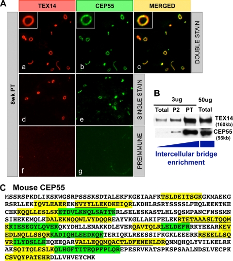
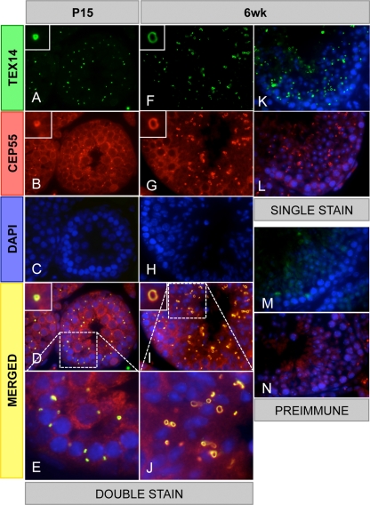
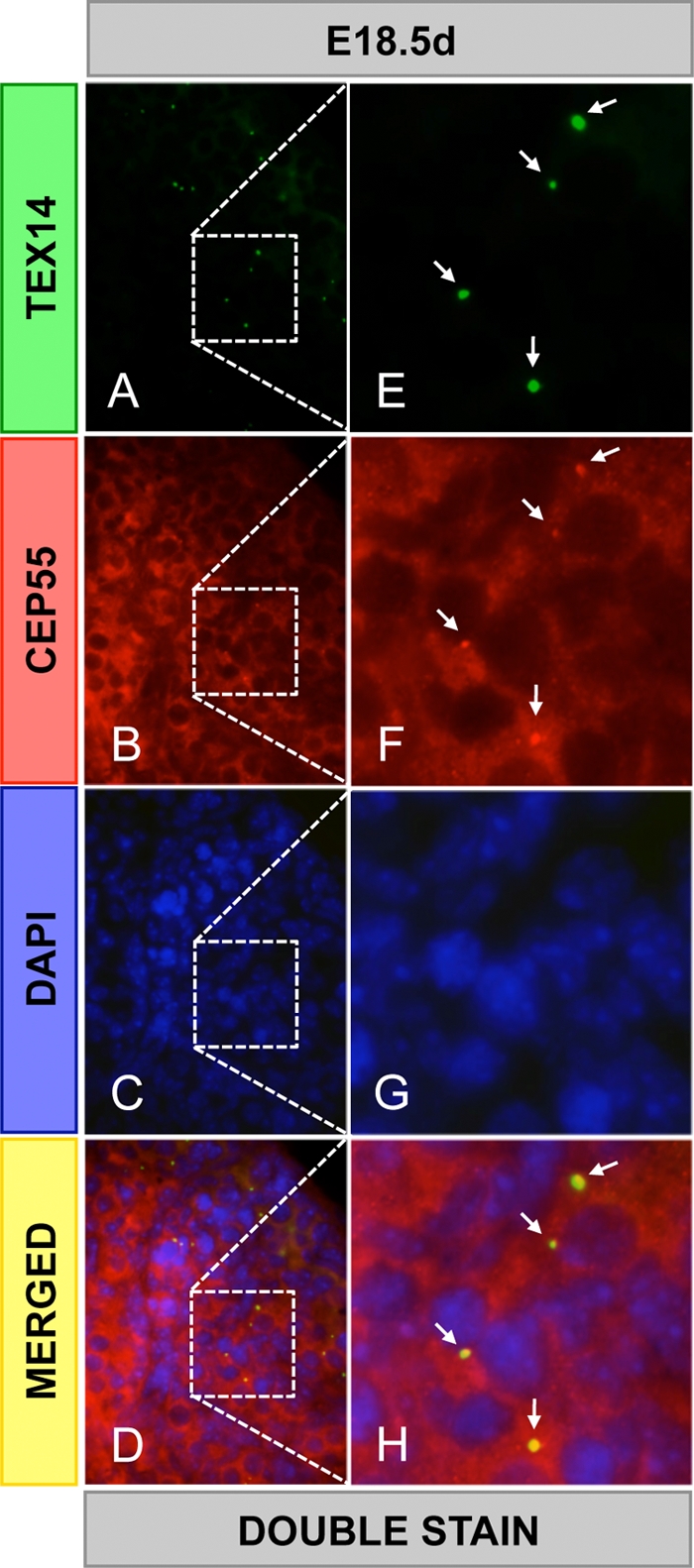
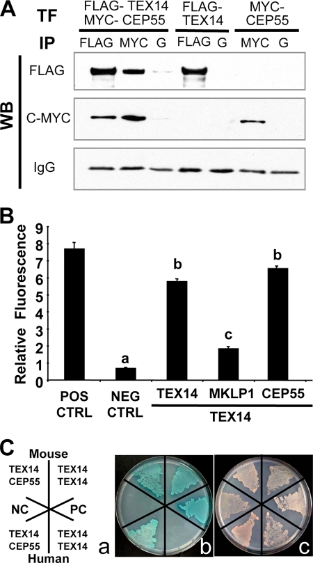
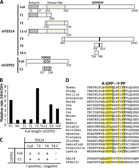
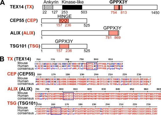
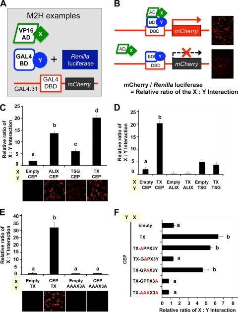
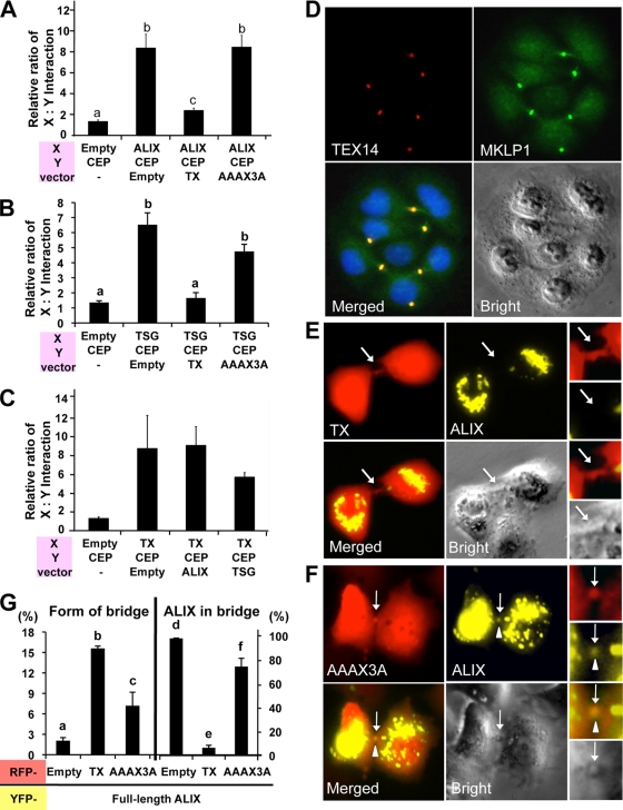
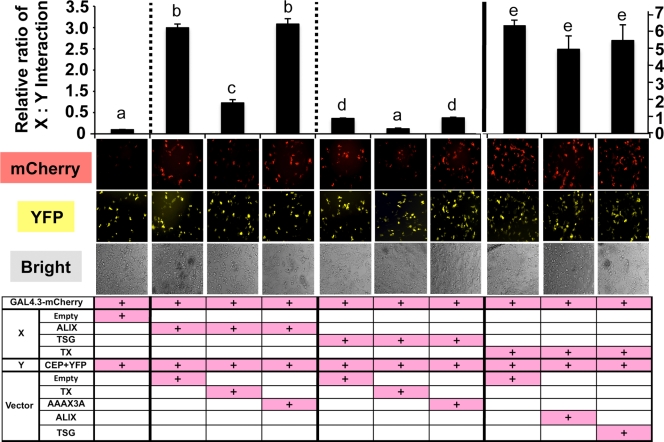
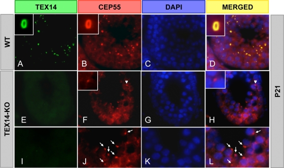
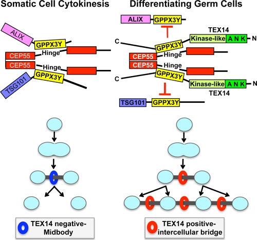
In somatic cells, abscission, the physical separation of daughter cells at the completion of cytokinesis, requires CEP55, ALIX, and TSG101. In contrast, cytokinesis is arrested prior to abscission in differentiating male germ cells that are interconnected by TEX14-positive intercellular bridges. We have previously shown that targeted deletion of TEX14 disrupts intercellular bridges in all germ cells and causes male sterility. Although these findings demonstrate that intercellular bridges are essential for spermatogenesis, it remains to be shown how TEX14 and other proteins come together to prevent abscission and form stable intercellular bridges. Using a biochemical enrichment of male germ cell intercellular bridges, we identified additional bridge proteins, including CEP55. Although CEP55 is highly expressed in testes at the RNA level, there is no report of the presence of CEP55 in germ cells. We show here that CEP55 becomes a stable component of the intercellular bridge and that an evolutionarily conserved GPPX3Y motif of TEX14 binds strongly to CEP55 to block similar GPPX3Y motifs of ALIX and TSG101 from interacting and localizing to the midbody. Thus, TEX14 prevents the completion of cytokinesis by altering the destiny of CEP55 from a nidus for abscission to an integral component of the intercellular bridge.


Figures

FIG. 1.
FIG. 1.
CEP55 is a component of the intercellular bridge. (A) Immunofluorescence was performed using custom-generated goat anti-mouse TEX14 and guinea pig anti-mouse CEP55 antibodies on isolated intercellular bridge preparations from an 8-week-old testis. The results from double staining (a to c), single staining (d and e), and preimmune using goat serum (f) or guinea pig serum (g) are shown as red, TEX14 or goat serum; green, CEP55 or guinea pig serum; and yellow, merged. (B) Western blot analysis of intercellular bridge enrichment using the anti-TEX14 and anti-CEP55 antibodies. (C) The 24 CEP55 peptides that were identified by proteomic analysis. The identified peptides are highlighted in yellow and green, with shorter overlapping peptides underlined.
FIG. 2.
FIG. 2.
CEP55 colocalizes with TEX14 in testes. Immunofluorescence was performed with rabbit anti-TEX14 and guinea pig anti-CEP55 antibodies in postnatal 15-day-old and 6-week-old mouse testes. The results from double staining (A to J), single staining (K and L), and preimmune using rabbit serum (M) or guinea pig serum (N) are shown; green, TEX14 or rabbit serum; red, CEP55 or guinea pig serum; blue, DAPI; yellow, merged. High-magnification images (E and J) are derived from the boxed regions of panels D and I.
FIG. 3.
FIG. 3.
CEP55 colocalizes with TEX14 in the ovary. Immunofluorescence was performed using goat anti-TEX14 and guinea pig anti-CEP55 antibodies in embryonic day 18.5 mouse ovaries. The results from double staining (A to H): green, TEX14; red, CEP55; blue, DAPI; and yellow, merged. High-magnification images (E to H) are derived from the boxed regions in panels A to D, respectively. Arrows, intercellular bridges.
FIG. 4.
FIG. 4.
TEX14 interacts with CEP55. (A) Immunoprecipitation (IP) of FLAG-TEX14 and/or MYC-CEP55 from HEK293T cells, followed by Western blot analysis with the antibodies as shown. Immunoprecipitation of protein G (lanes G) is the control. (B) Yeast two-hybrid analyses using vectors encoding full-length mouse TEX14, MKLP1, CEP55, and positive and negative controls. The relative ratios of TEX14-TEX14, TEX14-MKLP1, and TEX14-CEP55 interactions were determined by using an oxygen-biosensor system. (C) Yeast two-hybrid interactions of mouse and human full-length TEX14 and CEP55. (a) Yeast were stably transformed with vectors and plated for yeast two-hybrid analysis as depicted. SV40 T antigen with p53 and SV40 T antigen with lamin C were used as positive and negative controls, respectively. (b) Interactions between the two-hybrid proteins are evident by colony growth and blue color on selection plates. (c) A nonselection plate shows that all of the transformed yeast are capable of growing.
FIG. 5.
FIG. 5.
The region encoding the conserved TEX14 GPPX3Y motif interacts with the hinge region of CEP55. (A) The full-length and truncated regions of mouse TEX14 and CEP55 were cloned into the yeast two-hybrid vectors and used for the studies in panels B and C below. (B and C) Yeast two-hybrid oxygen biosensor between the full-length and truncated TEX14 proteins and the full-length CEP55 protein and/or selection plate analyses of the full-length and truncated TEX14 and CEP55 proteins. The terms “+” and “-” indicate positive and negative interactions, respectively. (D) Alignment of the GPPX3Y motif and flanking sequences from TEX14 orthologs and human ALIX and TSG101. Conserved amino acids are highlighted.
FIG. 6.
FIG. 6.
Essential motifs (A) and sequences (B) of TEX14, CEP55, ALIX, and TSG101. (A) The sizes and the domains/motif-containing regions of full-length TEX14, CEP55, ALIX, and TSG101 are shown. The domain regions highlighted in red are referred to as TX, CEP, ALIX, and TSG. (B) The corresponding amino acid sequences of these regions with the conserved consensus sequences in mouse and human are shown. These truncated proteins were used for the mammalian two-hybrid assays.
FIG. 7.
FIG. 7.
The GPPX3Y motif of TEX14 is essential for binding to the hinge region of CEP55. (A and B) Summary of the modified mammalian-two-hybrid assays (A) Three kinds of transfection vectors were made. One protein coding sequence (“X”) was fused to a transcriptional activation domain sequence (VP16-AD), and the other protein coding sequence (“Y”) was fused to a DNA-binding domain sequence (GAL4-BD). (B) When proteins “X” and “Y” interact, transcriptional activation of the mCherry gene occurs, which is detected as red fluorescence (B, top right). The GAL4-BD-Y vector expresses the Renilla reniformis luciferase, allowing for normalization of transfections. The relative interaction of protein X and Y is determined by the mCherry/Renilla reniformis luciferase ratio. (C to F) Mammalian-two-hybrid interactions of chimeric VP16-AD-X and GAL4-BD-Y proteins in transfected HEK293T cells is shown (see Fig. 6 and Table 2 for additional details).
FIG. 8.
FIG. 8.
The GPPX3Y motif of TEX14 inhibits the CEP55-ALIX and CEP55-TSG101 interactions and the entry of ALIX to the midbody, resulting in formation of stable intercellular bridges. (A to C) pcDNA3 vectors lacking an insert (Empty) or containing the truncated TEX14 (TX), ALIX (ALIX), TSG101(TSG), and TEX14 mutant (AAAX3A) were cotransfected into HEK293T cells along with VP16-AD-X, GAL4-BD-Y, and GAL4.31-mCherry vectors indicated at the bottom of each panel (see Fig. 6B and Table 2 for additional details). The relative ratios of the interactions of protein X and protein Y are shown. (D) Transfection of the full-length TEX14 vectors into HeLa cells. Immunofluorescence using goat anti-TEX14 and guinea pig anti-MKLP1 antibodies was performed: red, TEX14; green, MKLP1; blue, DAPI; and yellow, merged. (E and F) Cotransfection of pcDNA-YFP-full-length ALIX overexpression vector with pcDNA-mCherry-truncated GPPX3Y TEX14 (TX) (E) and TEX14 mutant AAAX3A (F) overexpression vectors into HeLa cells. The localization patterns of ALIX were microscopically examined for yellow fluorescence within a background of cells expressing TX or AAAX3A (red fluorescence). Arrow, midbody; arrowhead, ALIX. (G) Quantification of the experiment in panels E and F. The graphs were made by analyzing 1,000 double-positive cells with RFP and YFP from 11 to 13 separate experiments. The graphs show the percentage of the number of bridge containing cells/the number of double-positive RFP and YFP cells (left) and the number of ALIX localized in midbody/the number of RFP positive bridges in double-positive RFP and YFP cells (right).
FIG. 9.
FIG. 9.
The GPPX3Y-containing TEX14 region interacts with CEP55 much more strongly than the equivalent regions of ALIX and TSG101 and inhibits the CEP55-ALIX and CEP55-TSG101 interactions. The pcDNA3 vectors lacking an insert (Empty) or containing the truncated TEX14 (TX), ALIX (ALIX), TSG101(TSG), and TEX14 mutant (AAAX3A) were cotransfected into HEK293T cells, along with VP16-AD-X, GAL4-BD-Y, and GAL4.3.1-mCherry vectors indicated at the bottom of each panel. The GAL4-BD-Y vector, containing the Renilla luciferase sequence, was replaced by yellow fluorescent protein (YFP) sequence. YFP expression was used for normalization of transfections instead of Renilla luciferase. The relative interaction of proteins X and Y is determined by the mCherry/YFP ratio. The interactions of chimeric VP16-AD-X and GAL4-BD-Y proteins in transfected HEK293T cells are shown (see Fig. 6B and Table 2 for additional details).
FIG. 10.
FIG. 10.
TEX14 inhibits the completion of cytokinesis in the testis. Immunofluorescence of wild-type (WT) and TEX14 knockout (KO) testes from 3-week-old mice. TEX14 and CEP55 colocalize at the intercellular bridge in WT mouse testis (A to D). In TEX14 knockout testes (E to L), CEP55 localizes to midbodies (F and H, arrowhead) and abscissed midbodies (J and L, arrow). Green, TEX14; red, CEP55; blue, DAPI; yellow, merged.
FIG. 11.
FIG. 11.
Models for cytokinesis and intercellular bridge formation. (Left) Model of somatic cell abscission. CEP55 is essential in the recruitment of additional proteins (e.g., TSG101 and ALIX) that are required for abscission of the midbody (2, 3, 17). The regions of TSG101 and ALIX containing GPPX3Y motifs interact with the hinge region of CEP55 (14, 17) to complete cytokinesis. (Right) Model of the intercellular bridge in differentiating germ cells. The conserved GPPX3Y motif of TEX14 interacts strongly with the hinge region of CEP55 in differentiating germ cells to block CEP55 interactions with TSG101 and ALIX, resulting in formation of a stable intercellular bridge.

Similar articles

Cited by

References

    1. Braun, R. E., R. R. Behringer, J. J. Peschon, R. L. Brinster, and R. D. Palmiter. 1989. Genetically haploid spermatids are phenotypically diploid. Nature 337:373-376. - PubMed
    1. Carlton, J. G., M. Agromayor, and J. Martin-Serrano. 2008. Differential requirements for Alix and ESCRT-III in cytokinesis and HIV-1 release. Proc. Natl. Acad. Sci. U. S. A. 105:10541-10546. - PMC - PubMed
    1. Carlton, J. G., and J. Martin-Serrano. 2007. Parallels between cytokinesis and retroviral budding: a role for the ESCRT machinery. Science 316:1908-1912. - PubMed
    1. Carter, S. L., A. C. Eklund, I. S. Kohane, L. N. Harris, and Z. Szallasi. 2006. A signature of chromosomal instability inferred from gene expression profiles predicts clinical outcome in multiple human cancers. Nat. Genet. 38:1043-1048. - PubMed
    1. Dym, M., and D. W. Fawcett. 1971. Further observations on the numbers of spermatogonia, spermatocytes, and spermatids connected by intercellular bridges in the mammalian testis. Biol. Reprod. 4:195-215. - PubMed

Publication types

MeSH terms