. 2010 May;30(9):2251-63.
doi: 10.1128/MCB.01239-09. Epub 2010 Feb 22.

A comprehensive model that explains the regulation of phospholipase D2 activity by phosphorylation-dephosphorylation

Affiliations

A comprehensive model that explains the regulation of phospholipase D2 activity by phosphorylation-dephosphorylation

Karen M Henkels et al. Mol Cell Biol. 2010 May.

Abstract

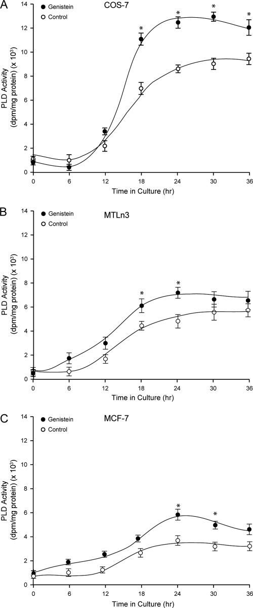
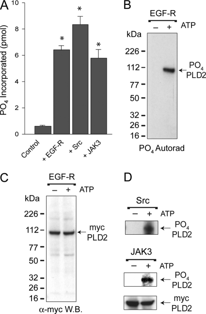
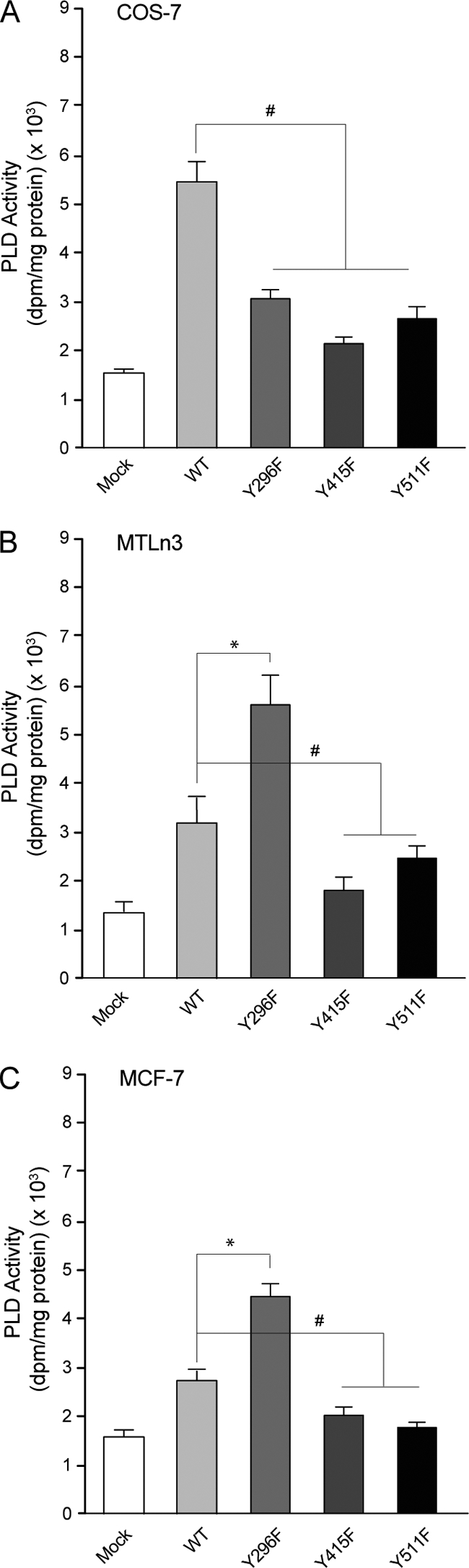
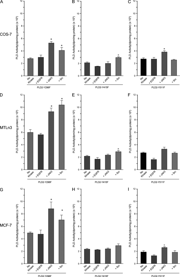
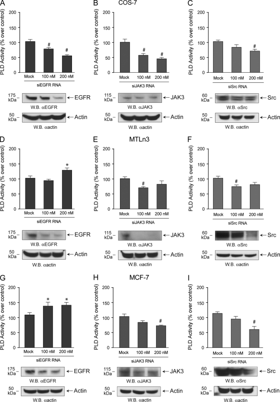
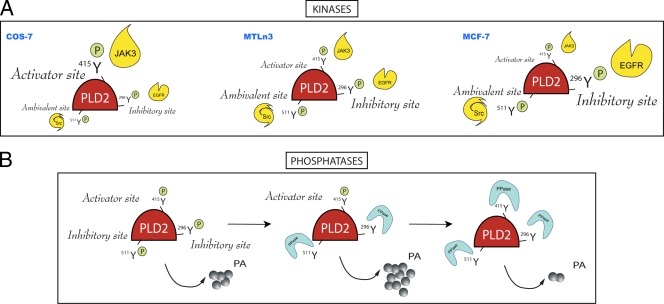
We report here that the enzymatic activity of phospholipase D2 (PLD2) is regulated by phosphorylation-dephosphorylation. Phosphatase treatment of PLD2-overexpressing cells showed a biphasic nature of changes in activity that indicated the existence of "activator" and "inhibitory" sites. We identified three kinases capable of phosphorylating PLD2 in vitro-epidermal growth factor receptor (EGFR), JAK3, and Src (with JAK3 reported for the first time in this study)-that phosphorylate an inhibitory, an activator, and an ambivalent (one that can yield either effect) site, respectively. Mass spectrometry analyses indicated the target of each of these kinases as Y(296) for EGFR, Y(415) for JAK3, and Y(511) for Src. The extent to which each site is activated or inhibited depends on the cell type considered. In COS-7, cells that show the highest level of PLD2 activity, the Y(415) is a prominent site, and JAK3 compensates the negative modulation by EGFR on Y(296). In MCF-7, cells that show the lowest level of PLD2 activity, the converse is the case, with Y(296) unable to compensate the positive modulation by Y(415). MTLn3, with medium to low levels of lipase activity, show an intermediate pattern of regulation but closer to MCF-7 than to COS-7 cells. The negative effect of EGFR on the two cancer cell lines MTLn3 and MCF-7 is further proven by RNA silencing experiments that yield COS-7 showing lower PLD2 activity, and MTLn3 and MCF-7 cells showing an elevated activity. MCF-7 is a cancer cell line derived from a low-aggressive/invasive form of breast cancer that has relatively low levels of PLD activity. We propose that PLD2 activity is low in the breast cancer cell line MCF-7 because it is kept downregulated by tyrosyl phosphorylation of Y(296) by EGFR kinase. Thus, phosphorylation of PLD2-Y(296) could be the signal for lowering the level of PLD2 activity in transformed cells with low invasive capabilities.


Figures

FIG. 1.
FIG. 1.
Expression of a PLD2 constitutively active in COS-7, MTLn3, and MCF-7 cells. Either COS-7 (A), MTLn3 (B), or MCF-7 (C) cells were transfected with pcDNA-mycPLD2-WT and divided into several sets. Each set was incubated with 0.1 μg/ml of genistein for the indicated times. Regardless of those variable lengths of time, all cells were allowed a full 36-h time for overexpression of PLD2. Overexpressed mycPLD2 was immunoprecipitated with 9E10 monoclonal antibody agarose matrix and assayed for lipase activity as indicated above. Enzyme reaction units in the y axes was calculated in relation to PLD2 protein, so comparison between the cell lines is possible. The results represent means ± the SEM of three independent experiments in duplicate. ANOVA tests were performed on the means and SEM of the different points and, when statistically significant differences (P < 0.05) were found, the points are denoted with an asterisk.
FIG. 2.
FIG. 2.
Biphasic regulation of PLD2 activity by tyrosine phosphatases in vitro. Either MCF-7, MTLn3, or COS-7 cells were transfected with pcDNA3.1-mycPLD2 for 36 h. Cell lysates were treated with the indicated concentrations of CD45 (A) or PTP1B (B) and incubated for 30 min at 30°C. Samples were then processed for PLD2 activity in PC8 liposomes and n-[3H]butanol. The results represent means ± the SEM of four independent experiments in duplicate. ANOVA tests were performed on the means and SEM of the different points comparing to controls and, when statistically significant differences (P < 0.05) were found, these points were denoted by “*” or “#”, respectively, for values above or below the basal level.
FIG. 3.
FIG. 3.
Differential effect of EGFR, Src, and JAK3 kinases on PLD2 activity. COS-7 (A and D), MTLn3 (B and E), or MCF-7 (C and F) cells (transfected with myc-PLD2 or mock transfected) were used for in vitro kinase reactions with EGFR, JAK3, or Src. In panels A to C, PLD2 was immunoprecipitated with anti-myc antibodies, and then the complex beads were mixed with the kinases. After the kinase reactions were completed, the products were taken for PLD activity assays. In panels D to F, whole lysates were first mixed with kinases and, after the kinase reactions were completed, PLD was immunoprecipitated with anti-myc antibodies. Finally, the complex beads were taken for PLD activity assays. In either case, the samples were split into two 50-μl aliquots for duplicate determinations in the PLD2 assay. The results represent means ± the SEM of four independent experiments in duplicate. *, differences between means (no-kinase versus kinase treatments) that were statistically significant (P < 0.05) as determined by ANOVA.
FIG. 4.
FIG. 4.
Investigation of the targets of phosphorylation. (A) Phosphate was incorporated into COS-7 PLD2 by the action of EGFR, Src, or JAK3 kinases in vitro. PLD2 α-myc-agarose-bound immunocomplexes were used for measurement of PO4 incorporation into PLD2. The results represent means ± the SEM of three independent experiments in duplicate. *, differences between means that were statistically significant (P < 0.05) as determined by ANOVA. (B) SDS-gel/autoradiograph of EGFR phosphorylated PLD2. Following kinase activations with or without [32P]ATP, proteins were electrophoresed and transferred onto PVDF membranes. Radioactive, phosphorylated PLD2 was detected by autoradiography. (C) Western blotting of EGFR phosphorylated PLD2. Nonradioactive, phosphorylated PLD2 was detected by using α-myc rabbit antibodies. (D) Representative autoradiograph of Src (first panel) or JAK3 (second panel). A representative Western blot of mycPLD2 (third panel) is also shown to indicate equal loading of the gels.
FIG. 5.
FIG. 5.
Detection of PLD2 phosphopeptides by MS. (A) HPLC. Lysates from COS-7 cells overexpressing PLD2 were immunoprecipitated with α-myc antibodies, phosphorylated, and run on SDS gels. Protein bands in the region of 105 ± 5 kDa were excised from the gels and trypsinized “in-gel.” The resulting tryptic peptides were separated by reversed-phase chromatography as shown. (B) The eluted samples from the HPLC column were analyzed by TOF-MS-ES. After this, an accurate mass analysis was performed to compare all parent ions to the theoretical tryptic digest of PLD2 using Biolynx software. A section of the spectrogram with an m/z of ∼1,100 for samples phosphorylated by Src is shown. (C) Identification of phosphorylated peptides. The compilation indicates m/z of samples that were positive for ions corresponding to phosphate adducts with double or triple charges and compared to the parent ions with the sequences of PLD2 (NCBI accession no. O14939). Also indicated are the sites most likely recognized for each of the kinases used in the present study (EGFR, Src, and JAK3).
FIG. 6.
FIG. 6.
Study of lipase activity for the three phosphorylation-incapable mutants. Phosphorylation-incapable PLD2-Y296F, PLD2-Y415F, and PLD2-Y511F, Y→F point mutants were generated by site-directed mutagenesis by using myc-pcDNA-PLD2-WT as a template. Each one of these mutants alone or the wild-type PLD2 (“WT”) or mock reagents was transfected into COS-7 (A), MTLn3 (B), or MCF-7 (C) cells. After 36 h, cells were harvested, and whole lysates were prepared from overexpressing cells and mixed in kinase buffer with either EGFR, JAK3, or Src kinases. After this, PLD2's were immunoprecipitated with α-myc antibodies. The PLD2-α-myc-agarose immunocomplexes were used for measurement of PLD activity in PC8 liposomes and n-[3H]butanol. * and #, differences between means, as determined by ANOVA, above and below, respectively, the wild-type levels that were statistically significant (P < 0.05).
FIG. 7.
FIG. 7.
Effect of exogenous kinases on the PLD activity of phosphorylation-deficient mutants. PLD2-Y296F (A, D, and G), PLD2-Y415F (B, E, and H), and PLD2-Y511F (C, F, and I) PLD2 mutants were transfected into COS-7 (A to C), MTLn3 (D to F), or MCF-7 (G to I) cells. After 36 h of expression, lysates were used for in vitro kinase reactions with EGFR, JAK3, or Src. After this, PLD2 was immunoprecipitated with α-myc antibodies, and samples were split into two 50-μl aliquots for duplicate determinations in the PLD2 assay. The results represent means ± the SEM of four independent experiments in duplicate. *, differences between means (no-kinase versus kinase treatments) that were statistically significant (P < 0.05) as determined by ANOVA.
FIG. 8.
FIG. 8.
Silencing endogenous kinases and status of PLD activity. EGFR, JAK3, or Src -siRNAs were transfected into COS-7 (A-C), MTLn3 (D-F) or MCF-7 (G-I) for 2 days. After this, duplicate cell samples were used to generate cell lysates for PLD activity assays and for the detection of protein expression by Western blotting. For the latter, blots were cut in half; the upper part (containing an ∼175-kDa region, an ∼115-kDA region, or an ∼60-kDA region) was probed with the appropriate anti-kinase antibodies, while the lower part (containing an ∼50-kDa region) was probed with anti-β-actin to ascertain equivalent protein loading.
FIG. 9.
FIG. 9.
Model highlighting the biological significance of the various findings from the present study. (A) Kinase action on PLD2. EGFR, JAK3, and Src are capable of phosphorylating PLD2 in vitro, and the targets are Y296 for EGFR, Y415 for JAK3, and Y511 for Src. EGFR phosphorylates an “inhibitory site,” JAK3 phosphorylates an “activator site,” and Src phosphorylates an “ambivalent site” (one that can yield either effect). The extent to which each site is activated or inhibited depends on the cell type considered. In COS-7 cells, which bear the highest level of PLD2 activity, the Y415 is a prominent site that, by being phosphorylated by JAK3, compensates for the negative modulation by EGFR on Y296. In MCF-7 cells, which show the lowest level of PLD2 activity, the converse is true, with Y296 being unable to compensate for the positive modulation by Y415. MTLn3 cells, with medium or low levels of lipase activity, show an intermediate pattern of regulation but one that is closer to that of MCF-7 cells than that of COS-7 cells. (B) Phosphatase action on PLD2. Low concentrations of phosphatases or phosphatases targeting Y511 or Y296 leave the enzyme regulated positively by the “activator” sites and thus with high levels of lipase activity. Conversely, with higher concentrations of phosphatases or with phosphatases targeting Y415 (the activator site), the enzyme loses activity to different extents based on how heavily the cell relies on activator or inhibitory sites. We propose that Y296, an inhibitory site in MCF-7 cancer cells, is the reason for the observed low lipase activity in them, and this underscores the importance of this site for regulation of PLD2.

Similar articles

Cited by

References

    1. Aguirre Ghiso, J. A., E. F. Farias, D. F. Alonso, C. Arregui, and E. Bal de Kier Joffe. 1997. A phospholipase D and protein kinase C inhibitor blocks the spreading of murine mammary adenocarcinoma cells altering f-actin and beta1-integrin point contact distribution. Int. J. Cancer 71:881-890. - PubMed
    1. Ahn, B. H., S. Y. Kim, E. H. Kim, K. S. Choi, T. K. Kwon, Y. H. Lee, J. S. Chang, M. S. Kim, Y.-H. Jo, and D. S. Min. 2003. Transmodulation between phospholipase D and c-Src enhances cell proliferation. Mol. Cell. Biol. 23:3103-3115. - PMC - PubMed
    1. Bae, C. D., D. S. Min, I. N. Fleming, and J. H. Exton. 1998. Determination of interaction sites on the small G protein RhoA for phospholipase D. J. Biol. Chem. 273:11596-11604. - PubMed
    1. Banno, Y., K. Ohguchi, N. Matsumoto, M. Koda, M. Ueda, A. Hara, I. Dikic, and Y. Nozawa. 2005. Implication of phospholipase D2 in oxidant-induced phosphoinositide 3-kinase signaling via Pyk2 activation in PC12 cells. J. Biol. Chem. 280:16319-16324. - PubMed
    1. Brown, H. A., S. Gutowski, C. R. Moomaw, C. Slaughter, and P. C. Sternweis. 1993. ADP-ribosylation factor, a small GTP-dependent regulatory protein, stimulates phospholipase D activity. Cell 75:1137-1144. - PubMed

Publication types

MeSH terms