. 2010 May;54(5):1988-99.
doi: 10.1128/AAC.01598-09. Epub 2010 Feb 22.

Discovery and characterization of inhibitors of Pseudomonas aeruginosa type III secretion

Affiliations

Discovery and characterization of inhibitors of Pseudomonas aeruginosa type III secretion

Daniel Aiello et al. Antimicrob Agents Chemother. 2010 May.

Abstract

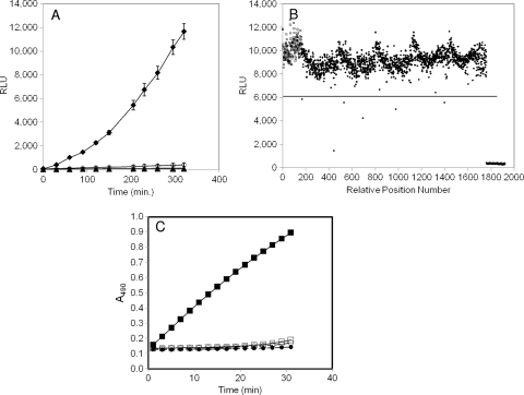
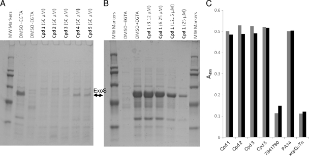
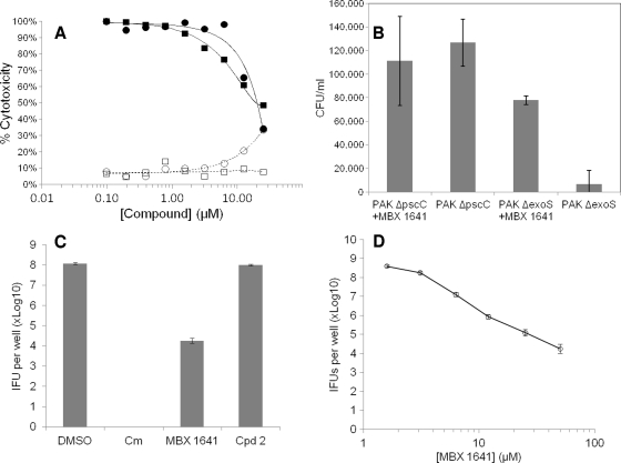
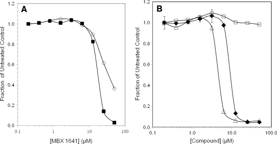
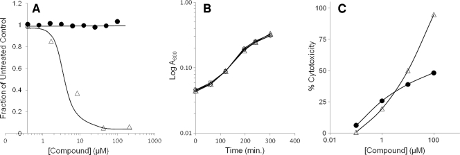
The type III secretion system (T3SS) is a clinically important virulence mechanism in Pseudomonas aeruginosa that secretes and translocates up to four protein toxin effectors into human cells, facilitating the establishment and dissemination of infections. To discover inhibitors of this important virulence mechanism, we developed two cellular reporter assays and applied them to a library of 80,000 compounds. The primary screen was based on the dependence of the transcription of T3SS operons on the T3SS-mediated secretion of a negative regulator and consisted of a transcriptional fusion of the Photorhabdus luminescens luxCDABE operon to the P. aeruginosa exoT effector gene. Secondary assays included direct measurements of the T3SS-mediated secretion of a P. aeruginosa ExoS effector-beta-lactamase fusion protein as well as the detection of the secretion of native ExoS by the sodium dodecyl sulfate-polyacrylamide gel electrophoresis (SDS-PAGE) analysis of culture supernatants. Five inhibitors in three chemical classes were demonstrated to inhibit type III secretion selectively with minimal cytotoxicity and with no effects on bacterial growth or on the type II-mediated secretion of elastase. These inhibitors also block the T3SS-mediated secretion of a YopE effector-beta-lactamase fusion protein from an attenuated Yersinia pestis strain. The most promising of the inhibitors is a phenoxyacetamide that also blocks the T3SS-mediated translocation of effectors into mammalian cells in culture. Preliminary studies of structure-activity relationships in this phenoxyacetamide series demonstrated a strict requirement for the R-enantiomer at its stereocenter and indicated tolerance for a variety of substituents on one of its two aromatic rings.


Figures

FIG. 1.
FIG. 1.
Characterization of bioluminescent and chromogenic reporter strains for identification of T3SS inhibitors. (A) Luminescence (in relative light units, RLU) from a chromosomal transcriptional fusion of exoT to the P. luminescens luxCDABE operon in wild-type (strain MDM852) or ΔpscC (strain MDM1355) P. aeruginosa PAO1 cells. Overnight cultures were diluted at time zero to A600 ∼ 0.025 and induced with 5 mM EGTA or left uninduced. RLU values were measured in 96-well opaque microplates throughout a 320-min time course. ⧫, MDM852 with EGTA; ⋄, MDM852 without EGTA; ▴, MDM1355 with EGTA; ▵, MDM1355 without EGTA. (B) Luminescence (RLU) from five 384-well microplates containing reporter strain MDM852 in a high-throughput screen for T3SS inhibitors. RLU values are shown at 300 min for 160 negative controls (□; fully induced by EGTA) in positions 1 to 160, for 160 positive controls (▴; no induction by EGTA) in positions 1,761 to 1,920, and for 1,600 samples (•) in positions 161 to 1,760. Six samples were designated hits because their RLU values displayed Z scores of >4 (i.e., >4 standard deviations below the average sample value, denoted as a horizontal line at 6,084 RLU). Compound 1 at position 443 was the most potent hit (Z score = 10) (Table 3). (C) Detection of ExoS′-βLA secretion from P. aeruginosa strains MDM973 (PAK) and MDM974 (PAK ΔpscC) carrying pUCP24GW-lacIQ-lacPO-exoS′-blaM, as measured by the hydrolysis of nitrocefin. A490 values are plotted versus time for MDM973 in the presence (▪) and absence (□) of 5 mM EGTA and for strain MDM974 in the presence (•) and absence (○) of 5 mM EGTA.
FIG. 2.
FIG. 2.
Evaluation of inhibition of type III and type II secretion in P. aeruginosa. P. aeruginosa ExoS-secreting strain PAKΔTY was grown under T3SS-inducing conditions (LB plus 5 mM EGTA) for 3 h in the presence of the indicated concentrations of compounds, and culture medium (1 ml) was concentrated in SDS-PAGE sample buffer, separated by SDS-12.5% PAGE, and stained with Coomassie blue. The positive control, DMSO plus EGTA, was treated with 5 mM EGTA but not inhibitors, and the negative control, DMSO without EGTA, was treated with neither EGTA nor inhibitors. The identity and molecular weights of protein markers are as follows: porcine myosin (200K), E. coli β-galactosidase (116K), rabbit muscle phosphorylase B (97K), bovine albumin (66K), ovalbumin (45K), and bovine carbonic anydrase (29K). (A) Secreted proteins from cells treated with EGTA and the five validated T3SS inhibitors (Table 3). The band corresponding to 49K ExoS is marked. (B) Secreted proteins from cells treated with EGTA and serial dilutions of T3SS inhibitor compound 1. (C) Effects of T3SS inhibitors on type II secretion of elastase. P. aeruginosa PA14 cells were grown in LB medium for 16 h in the presence of 50 μM of the indicated compounds. As controls, PA14 and PA14 xcpQ::Tn cells were grown in LB in the presence of the equivalent concentration of DMSO, and PA14 was grown in the presence of a type II secretion inhibitor (compound 7941790; Chembridge, Inc.). Culture medium corresponding to equivalent numbers of cells was harvested by centrifugation and incubated with shaking for 6 h with Congo Red-elastin. Digested soluble Congo Red was measured by the A495 in two independent assays and plotted (gray and black bars).
FIG. 3.
FIG. 3.
Inhibition of T3SS-mediated effects on mammalian cells in culture. (A) Concentration-dependent rescue of CHO cells from ExoU cytotoxicity by T3SS inhibitor MBX 1641. ExoU-secreting P. aeruginosa strain PAKΔSTYexoU was mixed with CHO cells at an MOI of 5 in the presence of MBX 1641 (•) or the known ExoU inhibitor pseudolipasin (▪) (27) at various concentrations as indicated. The percent cytotoxicity is calculated as the percentage of LDH released from cells intoxicated with P. aeruginosa with or without inhibitor compared to LDH released from intoxicated cells that were not treated with inhibitor. The effects of pseudolipasin (□) and MBX1641 (○) also are shown in the absence of P. aeruginosa cells to evaluate the inherent cytotoxicity of the compounds themselves. (B) T3SS inhibitor MBX 1641 relieves the ExoT block of the HeLa cell internalization of P. aeruginosa. HeLa cells were infected with P. aeruginosa PAK strains secreting ExoT (PAKΔS) or deficient in T3SS (PAKΔC) at an MOI of 10. MBX 1641 was added at 50 μM to half the wells containing each strain. After 2 h, cultures were treated with gentamicin (50 μg/ml) for an additional 2 h. HeLa cells were lysed with Triton, and serial dilutions were plated to determine the number of P. aeruginosa cells that had been protected from gentamicin by internalization. The CFU/ml of P. aeruginosa cells from lysed HeLa cells were determined in triplicate and plotted as the averages ± standard deviations. (C) MBX 1641 but not compound 2 inhibits the growth of C. trachomatis L2 cells in Hep-2 cells in culture. Confluent monolayer Hep-2 cells were infected with L2 at an MOI of 0.5 and treated with compounds at the indicated concentrations, followed by sonication and the measurement of IFUs on HeLa monolayers. Experiments were done in triplicate, and averages ± standard deviations are shown. Chloramphenicol (Cm) was used at 200 μg/ml as a positive control, and compound diluent (DMSO) was used as a negative control. (D) Concentration dependence of the inhibition of C. trachomatis L2 growth in Hep-2 cells by MBX 1641.
FIG. 4.
FIG. 4.
Inhibition of T3SS-mediated secretion of effector-β-lactamase fusion proteins. (A) Cells growing under T3SS-inducing conditions were treated for 3 h with MBX 1641, and secreted β-lactamase activity was measured by the cleavage of nitrocefin as the ΔA490/min. The rate of nitrocefin cleavage as a fraction of that of the untreated control is plotted versus the compound concentration. Bacterial species and effector βLA fusions were P. aeruginosa ExoS′-βLA (▪) and Y. pestis YopE-βLA (○). (B) Effects of MBX 1641 and its R- and S-enantiomers on ExoS′-βLA secretion from P. aeruginosa. Concentration dependence for MBX 1641 and its two stereo isomers, MBX 1684 and MBX 1686, were determined by the rate of nitrocefin cleavage by secreted ExoS′-βLA and calculated as the fraction of cleavage in the absence of inhibitor. Racemic mixture MBX 1641 (⧫), R-enantiomer MBX 1684 (Δ), and S-entantiomer MBX 1686 (□) are indicated.
FIG. 5.
FIG. 5.
Evaluation of the effects of MBX 1641 on bacterial and mammalian cell growth. (A) Determination of the MIC of MBX 1641 for P. aeruginosa. P. aeruginosa PAO1 cells were grown in the presence of the indicated concentrations of MBX 1641 (•) or tetracycline (▵) for 16 h in clear 96-well microplates, and the OD600 was determined. The OD600 as a fraction of that of DMSO-treated control cells is plotted. (B) Growth rate of P. aeruginosa cells treated with MBX 1641. P. aeruginosa PAO1 cells were grown in the presence of three different concentrations of MBX 1641 for 5 h in clear 96-well microplates, and the OD600 was measured periodically as indicated as a measure of cell density. MBX 1641 was present at 100 (□), 50 (•), or 25 μM (○), or cells were treated with an equivalent concentration (2%) of DMSO only (▵). (C) HeLa cell cytotoxicity of MBX 1641. HeLa cells were cultured in VP-SFM medium without serum in the presence of the indicated concentrations of MBX 1641 (•) or novobiocin (▵) for 3 days, and cytotoxicity was determined by the ability of remaining live cells to reduce a vital tetrazolium salt stain. Results are plotted as the percentage of cytotoxicity relative to levels for DMSO-treated and Triton X-100-lysed control cells.

Similar articles

Cited by

References

    1. Bradley, D. E. 1974. The adsorption of Pseudomonas aeruginosa pilus-dependent bacteriophages to a host mutant with nonretractile pili. Virology 58:149-163. - PubMed
    1. Carpino, L. A. 1993. 1-Hydroxy-7-azabenzotriazole. An efficient peptide coupling additive. J. Am. Chem. Soc. 115:4397-4398.
    1. Choi, K. H., A. Kumar, and H. P. Schweizer. 2006. A 10-min method for preparation of highly electrocompetent Pseudomonas aeruginosa cells: application for DNA fragment transfer between chromosomes and plasmid transformation. J. Microbiol. Methods 64:391-397. - PubMed
    1. Choi, K. H., and H. P. Schweizer. 2005. An improved method for rapid generation of unmarked Pseudomonas aeruginosa deletion mutants. BMC Microbiol. 5:30. - PMC - PubMed
    1. Clatworthy, A. E., E. Pierson, and D. T. Hung. 2007. Targeting virulence: a new paradigm for antimicrobial therapy. Nat. Chem. Biol. 3:541-548. - PubMed

Publication types

MeSH terms

LinkOut - more resources