Review
. 2010 Feb 22;188(4):453-61.
doi: 10.1083/jcb.200910105.

Automated microscopy for high-content RNAi screening

Affiliations
Review

Automated microscopy for high-content RNAi screening

Christian Conrad et al. J Cell Biol. .

Abstract

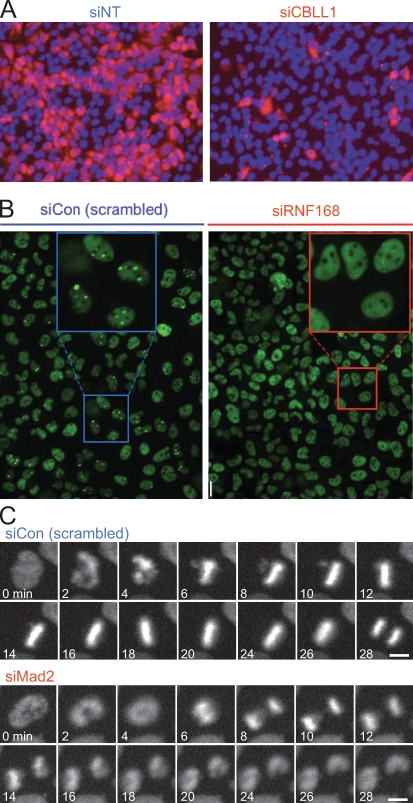
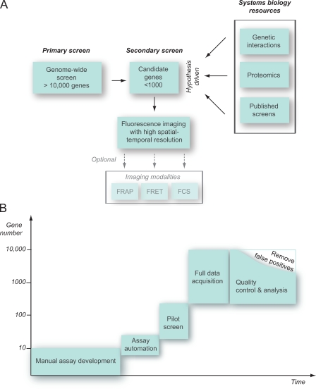
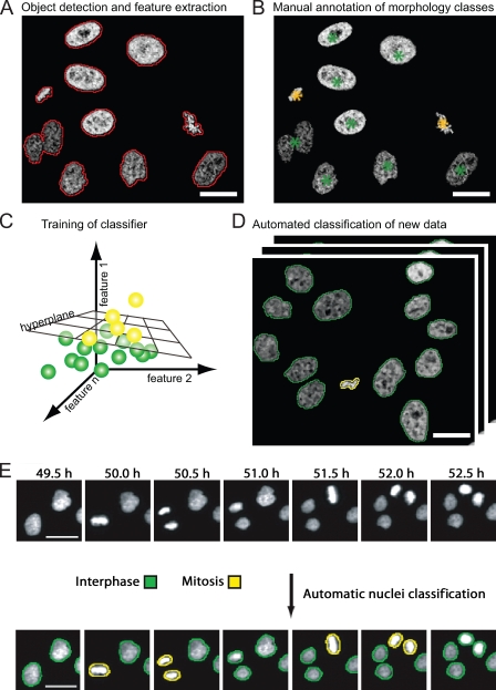
Fluorescence microscopy is one of the most powerful tools to investigate complex cellular processes such as cell division, cell motility, or intracellular trafficking. The availability of RNA interference (RNAi) technology and automated microscopy has opened the possibility to perform cellular imaging in functional genomics and other large-scale applications. Although imaging often dramatically increases the content of a screening assay, it poses new challenges to achieve accurate quantitative annotation and therefore needs to be carefully adjusted to the specific needs of individual screening applications. In this review, we discuss principles of assay design, large-scale RNAi, microscope automation, and computational data analysis. We highlight strategies for imaging-based RNAi screening adapted to different library and assay designs.


Figures

Figure 1.
Figure 1.
Examples for imaging-based assays. (A) Intensity-based assay. In this screen for human genes associated with West Nile virus infection, cell nuclei were labeled with DAPI (blue) and stained by immunofluorescence against a viral epitope (red). Genes that reduced the intensity of viral epitope staining were scored as hits. (left) Negative control (NT-siRNA). (right) Hit (CBLL1-siRNA) identified in the screen. This panel is reprinted with permission from Nature Publishing Group (Krishnan et al., 2008). (B) Morphology-based assay for genes functioning in the assembly of DNA double-strand repair foci. Cells were stained by immunofluorescence for 53BP1, a protein which localizes to spontaneously occurring DNA damage foci, visible as bright green spots (see negative control; left). In a hit obtained in this screen (siRNF168), 53BP1 failed to target to spots and was instead dispersed throughout the nucleoplasm. This panel is reprinted with permission from Elsevier (Doil et al., 2009). (C) Live imaging-based assay for mitotic timing. HeLa cells stably expressing chromatin marker H2B-mCherry (Steigemann et al., 2009) were imaged for 24 h on an incubated screening microscope (Schmitz and Gerlich, 2009). Mitotic phenotypes were detected based on the timing from nuclear breakdown (2 min in siCon) until anaphase (28 min in siCon). Cells depleted for the spindle checkpoint protein Mad2 prematurely enter anaphase (8 min). Images provided by D.W. Gerlich. Bars, 10 µm.
Figure 2.
Figure 2.
Screening strategies. (A) Secondary screening on candidate gene sets either derived from genome-wide primary screens (left) or derived based on a hypothesis and systems biology resources such as genetic interaction, proteomics, or bioinformatics data (right). The smaller size of perturbation conditions allows high-resolution spatial and temporal imaging or assays sampling protein mobility (for example, FRAP) or protein–protein interaction (for example, by fluorescence resonance energy transfer [FRET] or fluorescence correlation spectroscopy [FCS]). (B) Typical time line for implementation of an RNAi screen. Screening assays typically derive from a manual visual assay development, which aims at maximizing the discrimination between selected positive and negative controls. In the next step, the automation of sample preparation, data acquisition, storage, and analysis parameters has to be implemented and tested in a pilot screen. After the acquisition of the entire genome, quality control procedures reduce the number of validated hits.
Figure 3.
Figure 3.
Supervised machine learning for classification of cellular morphologies. (A) Detection of cells based on fluorescent chromatin label (core histone 2B fused to GFP; Kanda et al., 1998), as indicated by red contours. A set of quantitative texture and shape features is then extracted for each segmented object. (B) Manual annotation of morphology classes. In this example, interphase cell nuclei are annotated by green asterisks, and metaphase chromosome plates are annotated by yellow asterisks. (C) The hyperplane that optimally separates the two different morphology classes is automatically determined by the classification algorithm. The hyperplane can be defined by linear functions (as shown) or by radial base functions, depending on the classification algorithm. (D) The trained classifier can be applied to new data for automated classification of trained morphology classes. The yellow and green contours label interphase and metaphase morphology classes as shown in B. (E) Time-resolved assay for mitotic progression. Live HeLa cells expressing fluorescent core histone 2B (H2B-GFP) were imaged by automated time-lapse microscopy for 24 h using a 10× dry objective. Morphological classes were annotated by machine learning and pattern classification. This panel is reprinted with permission from Nature Publishing Group (Neumann et al., 2006). Bars, 20 µm.

Similar articles

Cited by

References

    1. Aye-Han N.N., Ni Q., Zhang J. 2009. Fluorescent biosensors for real-time tracking of post-translational modification dynamics. Curr. Opin. Chem. Biol. 13:392–397 10.1016/j.cbpa.2009.07.009 - DOI - PMC - PubMed
    1. Bakal C., Aach J., Church G., Perrimon N. 2007. Quantitative morphological signatures define local signaling networks regulating cell morphology. Science. 316:1753–1756 10.1126/science.1140324 - DOI - PubMed
    1. Bartz F., Kern L., Erz D., Zhu M., Gilbert D., Meinhof T., Wirkner U., Erfle H., Muckenthaler M., Pepperkok R., Runz H. 2009. Identification of cholesterol-regulating genes by targeted RNAi screening. Cell Metab. 10:63–75 10.1016/j.cmet.2009.05.009 - DOI - PubMed
    1. Birmingham A., Anderson E.M., Reynolds A., Ilsley-Tyree D., Leake D., Fedorov Y., Baskerville S., Maksimova E., Robinson K., Karpilow J., et al. 2006. 3′ UTR seed matches, but not overall identity, are associated with RNAi off-targets. Nat. Methods. 3:199–204 10.1038/nmeth854 - DOI - PubMed
    1. Birmingham A., Selfors L.M., Forster T., Wrobel D., Kennedy C.J., Shanks E., Santoyo-Lopez J., Dunican D.J., Long A., Kelleher D., et al. 2009. Statistical methods for analysis of high-throughput RNA interference screens. Nat. Methods. 6:569–575 10.1038/nmeth.1351 - DOI - PMC - PubMed

Publication types