Clinical Trial
. 2010 Mar 9;107(10):4758-63.
doi: 10.1073/pnas.0909074107. Epub 2010 Feb 22.

Functional specializations for music processing in the human newborn brain

Affiliations
Clinical Trial

Functional specializations for music processing in the human newborn brain

Daniela Perani et al. Proc Natl Acad Sci U S A. .

Abstract

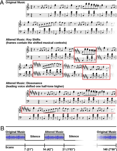
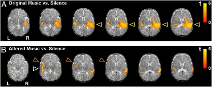
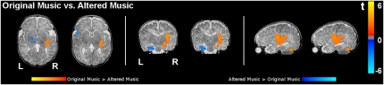
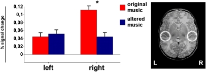
In adults, specific neural systems with right-hemispheric weighting are necessary to process pitch, melody, and harmony as well as structure and meaning emerging from musical sequences. It is not known to what extent the specialization of these systems results from long-term exposure to music or from neurobiological constraints. One way to address this question is to examine how these systems function at birth, when auditory experience is minimal. We used functional MRI to measure brain activity in 1- to 3-day-old newborns while they heard excerpts of Western tonal music and altered versions of the same excerpts. Altered versions either included changes of the tonal key or were permanently dissonant. Music evoked predominantly right-hemispheric activations in primary and higher order auditory cortex. During presentation of the altered excerpts, hemodynamic responses were significantly reduced in the right auditory cortex, and activations emerged in the left inferior frontal cortex and limbic structures. These results demonstrate that the infant brain shows a hemispheric specialization in processing music as early as the first postnatal hours. Results also indicate that the neural architecture underlying music processing in newborns is sensitive to changes in tonal key as well as to differences in consonance and dissonance.


Conflict of interest statement

The authors declare no conflict of interest.

Figures

Fig. 1.
Fig. 1.
Examples of stimuli and scanning paradigm. (A) Fragments illustrating the three sets of stimuli: original music, altered music: key shifts, and altered music: dissonance. (B) Experimental paradigm.
Fig. 2.
Fig. 2.
Activations elicited by the musical stimuli in newborns (n = 18, random effects group analyses, false discovery rate corrected; P < 0.0002 at the voxel level and P < 0.05 at the cluster level) overlaid over a T2-weighted image from a single newborn subject (note that the spatial resolution of the functional group data is lower compared with the anatomical image). (A) Mean activations for original music vs. silence are shown for six axial slices. Note the right-hemispheric predominance of temporal activation (yellow arrows). (B) Mean activations for altered music (key shifts and dissonance pooled) vs. silence. Note the left-hemispheric activation in the inferior frontal gyrus (orange arrows) and the reduced activation in the right temporal lobe (compared with the contrast of original music vs. silence, white arrow). (Details are provided in Materials and Methods.)
Fig. 3.
Fig. 3.
Direct contrast of original music vs. altered music in healthy newborns (n = 18, random effects group analysis; P < 0.05 at the voxel level, uncorrected) overlaid on a T2-weighted image from a single newborn (note that the spatial resolution of the functional group data is lower compared with the anatomical image). Regions more active for original music are shown in orange/yellow, and regions more active for altered music are shown in blue. Two axial slices show a stronger activation of the left inferior frontal gyrus in response to altered music. The slices also show a stronger activation of (posterior) auditory cortex in response to original music. The two coronal slices show activation of the left amygdala-hippocampal complex (and of the ventral striatum) for altered music and activation of the right amygdala-hippocampal complex for original music. The two sagittal slices show the larger right superior temporal activation for original music.
Fig. 4.
Fig. 4.
ROI analysis. Changes in activation for original music and altered music (key shifts and dissonance pooled) in primary and secondary auditory cortices as measured within spherical ROIs. (Right) ROIs on a T2-weighted image of a single newborn subject. The histograms show the percent signal change measured in each ROI during each of the two stimulus types (original music and altered music). Error bars indicate SEM.

Similar articles

Cited by

References

    1. Peretz I, Zatorre RJ. Brain organization for music processing. Annu Rev Psychol. 2005;56:89–114. - PubMed
    1. Gaab N, Gaser C, Schlaug G. Improvement-related functional plasticity following pitch memory training. NeuroImage. 2006;31:255–263. - PubMed
    1. Münte TF, Altenmüller E, Jäncke L. The musician's brain as a model of neuroplasticity. Nat Rev Neurosci. 2002;3:473–478. - PubMed
    1. Zatorre RJ, Chen JL, Penhune VB. When the brain plays music: Auditory-motor interactions in music perception and production. Nat Rev Neurosci. 2007;8:547–558. - PubMed
    1. Molnar-Szakacs I, Overy K. Music and mirror neurons: From motion to ‘e’ motion. Soc Cogn Affect Neurosci. 2006;1:235–241. - PMC - PubMed

Publication types