. 2010 Mar 9;107(10):4601-6.
doi: 10.1073/pnas.0909501107. Epub 2010 Feb 22.

Follistatin-like-1, a diffusible mesenchymal factor determines the fate of epithelium

Affiliations

Follistatin-like-1, a diffusible mesenchymal factor determines the fate of epithelium

Tomohiro Umezu et al. Proc Natl Acad Sci U S A. .

Abstract

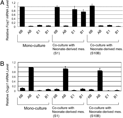
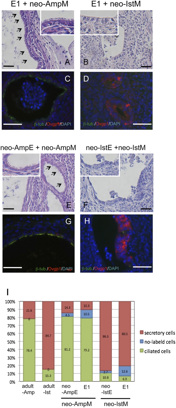
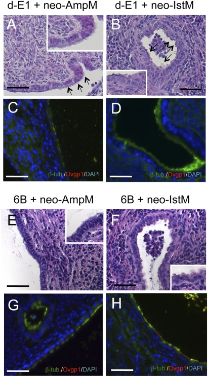
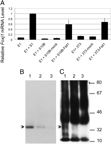
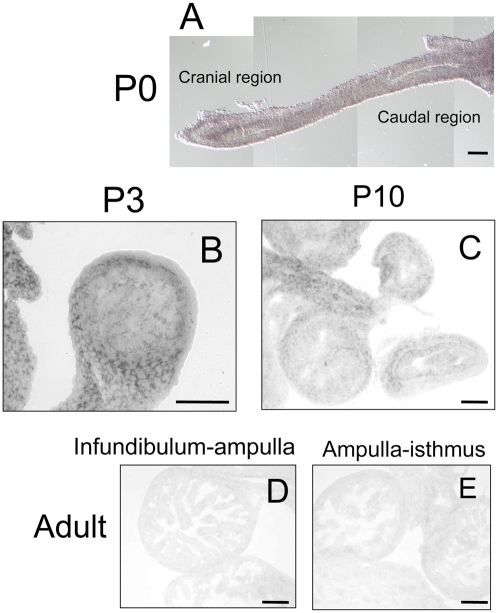
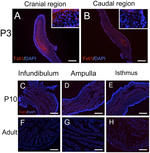
Mesenchyme is generally believed to play critical roles in "secondary induction" during organogenesis. Because of the complexity of tissue interactions in secondary inductions, however, little is known about the precise mechanisms at the cellular and molecular levels. We have demonstrated that, in mouse oviductal development, the mesenchyme determines the fate of undetermined epithelial cells to become secretory or cilial cells. We have established a model for studying secondary induction by establishing clonal epithelial and mesenchymal cell lines from perinatal p53(-/-) mouse oviducts. The signal sequence trap method collected candidate molecules secreted from mesenchymal cell lines. Naive epithelial cells exposed to Follistatin-like-1 (Fstl1), one of the candidates, became irreversibly committed to expressing a cilial epithelial marker and differentiated into ciliated cells. We concluded that Fstl1 is one of the mesenchymal factors determining oviductal epithelial cell fate. This is a unique demonstration that the determination of epithelial cell fate is induced by a single diffusible factor.


Conflict of interest statement

The authors declare no conflict of interest.

Figures

Fig. 1.
Fig. 1.
Marker expressions of epithelial cell lines cocultured with mesenchymal cell lines. Epithelial cell lines and mesenchymal cell lines were cocultured for 24 h on a culture insert and on a dish, respectively. (A) Foxj1 (ciliogenesis marker) expression of epithelial cell lines was analyzed by real-time RT–PCR. In monoculture, cilial epithelial cells (6B, positive control) had Foxj1 expression, but secretory epithelial cells (AB, negative control) and E1 and B1 epithelial cells had no Foxj1 expression. In coculture with S1 mesenchymal cells, E1 cells and B1 cells expressed Foxj1 to the same level as the positive control did. (B) Ovgp1 expression of epithelial cell lines analyzed by real-time RT–PCR. Secretory epithelial cells (AB) had Ovgp1 expression in monoculture (positive control). Error bars show the SD (n = 3).
Fig. 2.
Fig. 2.
Histology and immunohistochemistry of recombinant tissues. Neo-AmpM (mesenchyme of ampulla) and neo-IstM (mesenchyme of isthmus) of P3 oviducts were recombined with E1 epithelial cells and grown under the kidney capsule for 4 weeks. As control, neo-AmpE and neo-IstE (epithelium of P3 oviducts) were recombined with neo-AmpM and neo-IstM, respectively. (Scale bars: 100 μm.) (A) E1 epithelial cells combined with neo-AmpM developed epithelia with numerous cilia (arrows). (B) E1 epithelial cells combined with neo-IstM developed epithelia of simple columnar cells without cilia. (C) Double immunostaining for β-tubulin IV (green) and Ovgp1 (red). β-Tubulin IV-positive cells occupied the epithelium. (D) Double immunostaining for β-tubulin IV and Ovgp1 of reconstructed oviducts with E1 epithelial cells and neo-IstM. Ovgp1-positive cells occupied the epithelium. (E) Neo-AmpE was recombined with neo-AmpM. Numerous cilia were observed at the surface of the epithelium (arrows). (F) Neo-IstE was recombined with neo-IstM. The epithelium was occupied mainly by simple columnar cells without cilia. Insets in A, B, E, and F are partial enlargements (×4). (G) Double immunostaining for β-tubulin IV and Ovgp1 of reconstructed oviducts with neo-AmpE and neo-AmpM. β-Tubulin IV-positive cells occupied the epithelium. (H) Double immunostaining for β-tubulin IV and Ovgp1 of reconstructed oviducts with neo-IstE and neo-IstM. Ovgp1-positive cells occupied the epithelium. (I) Percentage of β-tubulin IV-positive cells and Ovgp1-positive cells in epithelia of adult oviducts and reconstructed oviducts. To determine the ratio of ciliated epithelial cells (β-tubulin IV-positive) and secretory epithelial cells (Ovgp1-positive), the numbers of β-tubulin IV-positive epithelial cells, Ovgp1-positive epithelial cells, and double-negative epithelial cells were counted on the screen in three frames for each specimen (>400 total epithelial cells). Results were based on an analysis of 8–24 tissue recombinants per group. In ampulla of adult oviducts, epithelia had β-tubulin IV-positive cells (78.4 ± 6.2%) and Ovgp1-positive cells (21.6 ± 3.1%) (n = 3). In the isthmus of adult oviducts, epithelia had β-tubulin IV-positive cells (15.3 ± 1.4%) and Ovgp1-positive cells (84.7 ± 5.8%) (n = 3). In recombinants of neo-AmpE and neo-AmpM, epithelia had β-tubulin IV-positive cells (81.2 ± 7.3%) and Ovgp1-positive cells (14.3 ± 2.6%). Double-negative cells were 4.5 ± 1.3% (n = 8). In recombinants of E1 cells and neo-AmpM, epithelia had β-tubulin IV-positive cells (79.2 ± 7.7%) and Ovgp1-positive cells (10.3 ± 2.6%). Double-negative cells were 10.5 ± 1.6% (n = 12). In recombinants of neo-IstE and neo-IstM, epithelia had β-tubulin IV-positive cells (10.8 ± 1.2%) and Ovgp1-positive cells (86.5 ± 8.6%). Double-negative cells were 2.7 ± 0.3% (n = 8). In recombinants of E1 epithelial cells and neo-IstM, epithelia had β-tubulin IV-positive cells (6.9 ± 1.1%) and Ovgp1-positive cells (80.5 ± 5.5%). Double-negative cells were 12.6 ± 2.7% (n = 12).
Fig. 3.
Fig. 3.
Fate determination is irreversible. Recombined tissues were implanted under the kidney capsule for 4 weeks. d-E1 epithelial cells were combined with neo-AmpM (P3 mesenchyme of ampulla) and neo-IstM (P3 mesenchyme of isthmus). As control, 6B cells (an adult epithelial line) were combined with neo-AmpM and neo-IstM. (Scale bars: 100 μm.) (A) d-E1 epithelial cells combined with neo-AmpM developed epithelia with numerous cilia at the surface (arrows). (B) E1 epithelial cells combined with neo-IstM also developed epithelia with numerous cilia at the surface (arrows). (C) Double immunostaining for β-tubulin IV (green) and Ovgp1 (red) of reconstructed oviducts with d-E1 epithelial cells and neo-AmpM. β-Tubulin IV-positive cells occupied the epithelium, and no Ovgp1-positive cells were observed. (D) Double immunostaining for β-tubulin IV and Ovgp1 of reconstructed oviducts with d-E1 epithelial cells and neo-IstM. β-Tubulin IV-positive cells occupied the epithelium and no Ovgp1-positive cells were observed. (E) 6B epithelial cells combined with neo-AmpM developed epithelia with numerous cilia at the surface. (F) 6B cells (an adult epithelial line) combined with neo-IstM developed epithelia with numerous cilia at the surface as observed when combined with neo-AmpM. Insets in A, B, E, and F are partial enlargements (×4). (G) Double immunostaining for β-tubulin IV and Ovgp1 of reconstructed oviducts with 6B epithelial cells and neo-AmpM. β-Tubulin IV-positive cells occupied the epithelium. (H) Double immunostaining for β-tubulin IV and Ovgp1 of reconstructed oviducts with 6B epithelial cells and neo-IstM. β-Tubulin IV-positive cells occupied the epithelium as observed when combined with neo-AmpM.
Fig. 4.
Fig. 4.
Coculture of undetermined epithelial cells and Fstl1-overexpressing mesenchymal cells. (A) E1 epithelial cells were cocultured with Fstl1 overexpressing cells for 24 h and subjected to real-time RT–PCR analysis of Foxj1 (ciliogenesis marker) expression. Error bars show the standard deviation (n = 3). (B) Western blot analysis of Fstl1 in lysates of 3T3-Fstl1 cells (lane 1), S1 cells (lane 2), and S10B cells (lane 3). Fstl1 protein was detected at 34 kDa in lysates of 3T3-Fstl1 cells and S1 mesenchymal cells (lanes 1 and 2), but not in the sample of S10B mesenchymal cells (lane 3). (C) Immunoprecipitation analysis detected Fstl1 protein at 34 kDa in media conditioned by 3T3-Fstl1 and S1 cells (lanes 1 and 2), but not in medium conditioned by S10B cells (lane 3).
Fig. 5.
Fig. 5.
Localization of FSTL1 mRNA in oviducts. In situ hybridization was performed to localize the expression of Fstl1 in oviducts. (Scale bars: 100 μm.) (A) Signals of Fstl were detected in the cranial part of P0 oviducts. (B) Signals of Fstl were strongly and specifically detected in the mesenchymal compartment of P3 oviducts. (C) Signals of Fstl became weaker in P4 oviducts. (D and E) Signals of Fstl were undetected in any region of adult oviducts.
Fig. 6.
Fig. 6.
Localization of FSTL1 in oviducts. (A and B) Stronger immunoreaction for Fstl1 (red) was observed in the cranial region than in the caudal region, and the reaction was detected mainly in mesenchymal cells of P3 oviducts. The sections were counterstained with DAPI (blue). Insets in A and B are partial enlargements (5×). (CE) Immunoreaction was not detected in the infundibulum, ampulla, and isthmus of P10 oviducts. (FH) Immunoreaction was not detected in the infundibulum, ampulla, and isthmus of adult oviducts.

Similar articles

Cited by

References

    1. Spemann H. Embryonic development and induction. New Haven, CT: Yale University Press; 1938.
    1. De Robertis EM. Spemann's organizer and self-regulation in amphibian embryos. Nat Rev Mol Cell Biol. 2006;7:296–302. - PMC - PubMed
    1. Ruiz i Altaba A. Induction and axial patterning of the neural plate: Planar and vertical signals. J Neurobiol. 1993;24:1276–1304. - PubMed
    1. Kuroda H, Wessely O, De Robertis EM. Neural induction in Xenopus: Requirement for ectodermal and endomesodermal signals via Chordin, Noggin, Catenin, and Cerberus. PLoS Biol. 2004;2 - PMC - PubMed
    1. Grobstein C. Trans-filter induction of tubules in mouse metanephrogenic mesenchyme. Exp Cell Res. 1956;10:424–440. - PubMed

Publication types

MeSH terms

LinkOut - more resources