

Early detection of superficial squamous cell carcinoma in the head and neck region and esophagus by narrow band imaging: a multicenter randomized controlled trial
- PMID: 20177025
- PMCID: PMC2849774
- DOI: 10.1200/JCO.2009.25.4680
Early detection of superficial squamous cell carcinoma in the head and neck region and esophagus by narrow band imaging: a multicenter randomized controlled trial
Abstract
Purpose: Most of the esophageal squamous cell carcinomas (ESCCs) and cancers of the head and neck (H&N) region are diagnosed at later stages. To achieve better survival, early detection is necessary. We compared the real-time diagnostic yield of superficial cancer in these regions between conventional white light imaging (WLI) and narrow band imaging (NBI) in high-risk patients.
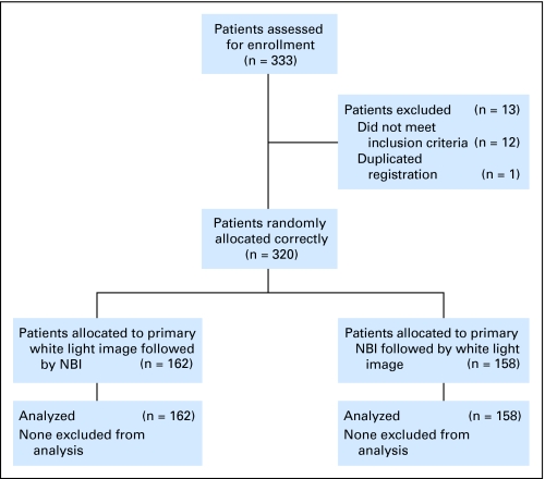
Patients and methods: In a multicenter, prospective, randomized controlled trial, 320 patients with ESCC were randomly assigned to primary WLI followed by NBI (n = 162) or primary NBI followed by WLI (n = 158) in a back-to-back fashion. The primary aim was to compare the real-time detection rates of superficial cancer in the H&N region and the esophagus between WLI and NBI. The secondary aim was to evaluate the diagnostic accuracy of these techniques.
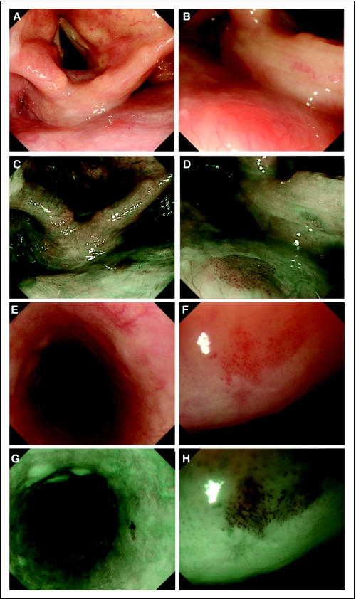
Results: NBI detected superficial cancer more frequently than did WLI in both the H&N region and the esophagus (100% v 8%, P < .001; 97% v 55%, P < .001, respectively). The sensitivity of NBI for diagnosis of superficial cancer was 100% and 97.2% in the H&N region and the esophagus, respectively. The accuracy of NBI for diagnosis of superficial cancer was 86.7% and 88.9% in these regions, respectively. The sensitivity and accuracy were significantly higher using NBI than WLI in both regions (P < .001 and P = .02 for the H&N region; P < .001 for both measures for the esophagus, respectively).
Conclusion: NBI could be the standard examination for the early detection of superficial cancer in the H&N region and the esophagus.
Conflict of interest statement
Authors' disclosures of potential conflicts of interest and author contributions are found at the end of this article.
Figures
References
-
- Parkin DM, Bray F, Ferlay J, et al. Global cancer statistics, 2002. CA Cancer J Clin. 2005;55:74–108. - PubMed
-
- Mori M, Adachi Y, Matsushima T, et al. Lugol staining pattern and histology of esophageal lesions. Am J Gastroenterol. 1993;88:701–705. - PubMed
-
- Inoue H, Rey JF, Lightdale C. Lugol chromoendoscopy for esophageal squamous cell cancer. Endoscopy. 2001;33:75–79. - PubMed
-
- Muto M, Hironaka S, Nakane M, et al. Association of multiple Lugol-voiding lesions with synchronous and metachronous esophageal squamous cell carcinoma in patients with head and neck cancer. Gastrointest Endosc. 2002;56:517–521. - PubMed
-
- Gono K, Yamazaki K, Doguchi N, et al. Endoscopic observation of tissue by narrow band illumination. Opt Rev. 2003;10:211–215.
Publication types
MeSH terms
LinkOut - more resources
Full Text Sources
Other Literature Sources
Medical
