. 2010 Apr 16;285(16):12445-53.
doi: 10.1074/jbc.M109.096735. Epub 2010 Feb 20.

The endoplasmic reticulum Grp170 acts as a nucleotide exchange factor of Hsp70 via a mechanism similar to that of the cytosolic Hsp110

Affiliations

The endoplasmic reticulum Grp170 acts as a nucleotide exchange factor of Hsp70 via a mechanism similar to that of the cytosolic Hsp110

Claes Andréasson et al. J Biol Chem. .

Abstract

Grp170 and Hsp110 proteins constitute two evolutionary distinct branches of the Hsp70 family that share the ability to function as nucleotide exchange factors (NEFs) for canonical Hsp70s. Although the NEF mechanism of the cytoplasmic Hsp110s is well understood, little is known regarding the mechanism used by Grp170s in the endoplasmic reticulum. In this study, we compare the yeast Grp170 Lhs1 with the yeast Hsp110 Sse1. We find that residues important for Sse1 NEF activity are conserved in Lhs1 and that mutations in these residues in Lhs1 compromise NEF activity. As previously reported for Sse1, Lhs1 requires ATP to trigger nucleotide exchange in its cognate Hsp70 partner Kar2. Using site-specific cross-linking, we show that the nucleotide-binding domain (NBD) of Lhs1 interacts with the NBD of Kar2 face to face, and that Lhs1 contacts the side of the Kar2 NBD via its protruding C-terminal alpha-helical domain. To directly address the mechanism of nucleotide exchange, we have compared the hydrogen-exchange characteristics of a yeast Hsp70 NBD (Ssa1) in complex with either Sse1 or Lhs1. We find that Lhs1 and Sse1 induce very similar changes in the conformational dynamics in the Hsp70. Thus, our findings demonstrate that despite some differences between Hsp110 and Grp170 proteins, they use a similar mechanism to trigger nucleotide exchange.


Figures
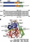
FIGURE 1.
FIGURE 1.
Lhs1 shares subdomain organization with Sse1. A, model for Lhs1 domain (NBD, SBD) and subdomain organization compiled from sequence alignment, local homology searches, and secondary structure prediction of Sse1 and fungal Lhs1 homologues. Indicated are the signal sequence (ss, amino acid residues 1–20), NBD (21–433, blue box), SBDβ sheet 1–7 (441–547, brown box), SBDβ loop (L, 548–564, yellow box), SBDβ sheet 8 (565–577, brown box), SBDα helix B (582–627, green box), SBDα helix C (635–650, green box), and SBDα helix DE (663–706, green box). Lhs1 has an extended C terminus with no significant homology to Sse1-(708–881). Homologous domains and subdomains of Sse1 are according to Ref. . Inset displays a sequence alignment of SBDα helix B from four fungal Lhs1 (S. cerevisiae, SC; K. lactis, KL; A. gossypii, Ag; C. albicans, Ca) and Sse1. Lhs1 Asn608 and Glu611 (boxed) exhibit sequence conservation with corresponding Sse1 residues (Asn572 and Glu575) that contact the Hsp70 NBD and are required for NEF activity (8). B, structure of a complex between Sse1 and the Hsp70 NBD (8). Sse1 is colored as in A and the Hsp70 NBD is colored red. Sse1 residues Thr280/Asn281, Thr365/Asn367, and Asn572/Glu575 that contact the Hsp70 NBD are marked (yellow). Residues used for pairwise cross-linking in Fig. 4 are marked (yellow, dotted lines) and corresponding positions in Lhs1/Sse1 and Hsp70/Kar2/Ssa1 are as indicated (table).
FIGURE 2.
FIGURE 2.
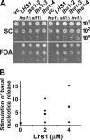
Residues conserved between Lhs1 and Sse1 are important for NEF activity. A, 10-fold serial dilutions of yeast strains CAY1171 (lhs1Δ sil1Δ) and CAY1172 (lhs1Δ sil1Δ ire1Δ) transformed to His+ using a vector control (vc), pCA715 (LHS1), or pCA715 carrying lhs1–2, lhs1–3, or lhs1–4 alleles. Cells were spotted onto control synthetic complete medium (SC) and medium containing FOA to counterselect against pCA716 (URA3 LHS1) that is present in the strains. Plates were incubated for 48 h at 30 °C. B, fold-stimulation of the basal rate of MABA-ADP dissociation from Kar2 NBD (2 μm) at the indicated concentrations of Lhs1 (♦) and derived mutants Lhs1–2 (■), Lhs1–3 (▲), and Lhs1–4 (●). Note that the data points for 4 μm Lhs1–2 and Lhs1–4 overlap. Proteins were preincubated with 3 mm ATP before measuring the release rates at 30 °C with a stopped flow instrument.
FIGURE 3.
FIGURE 3.
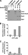
Lhs1 requires ATP to associate with Kar2 and trigger nucleotide exchange. A, Lhs1 and His6-Smt3-Kar2 were incubated in the presence and absence of ATP and loaded onto cobalt-NTA matrix. After washing, bound protein was eluted by proteolytic cleavage of the His6-Smt3 using Ulp1 protease and analyzed on SDS-PAGE. B, fold-stimulation of the basal rate of MABA-ADP dissociation from Kar2 NBD (2 μm) induced by Lhs1 (2 μm) preincubated at the indicated concentrations of ATP. The release rates were measured with a stopped flow instrument. C, ATP-dependent interaction between Lhs1–2, Lhs1–3, and Lhs1–4 and Kar2. Quantitation of pulldown assays (see A) given as percent of wild-type Lhs1 bound to the Kar2 NBD. Error bars indicate the standard error (n = 4).
FIGURE 4.
FIGURE 4.
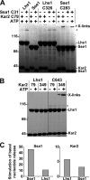
Lhs1 and Sse1 interact with Hsp70s in a similar manner. A, Lhs1, Sse1, or derivatives carrying introduced cysteine residues (Lhs1-C326 and Sse2-C283) were preincubated in the presence or absence of 1 mm ATP with the NBDs of Kar2 and Ssa1 carrying introduced cysteine residues (Kar2-C79 and Ssa1-C31). After cross-linking with BMH, cross-linked products were detected by SDS-PAGE analysis. The asterisk (*) refers to background protein bands that are not products of cross-linking. B, control Lhs1 and Lhs1 carrying a cysteine at position 643 (C643) was tested for cross-linking (as in A) to Kar2 NBDs carrying cysteines in place of residues 79 or 349. The asterisk (*) refers to background protein bands that are not products of cross-linking. C, fold-stimulation of the basal rate of MABA-ADP dissociation from the Kar2 and Ssa1 NBDs (0.5 μm) induced by Lhs1 or Sse1 (1 μm). Lhs1 and Sse1 were preincubated with 0.5 mm ATP before measuring the release rates with a stopped flow instrument.
FIGURE 5.
FIGURE 5.
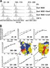
HX properties of the Ssa1 NBD in complex with Lhs1. A, mass spectra of representative peptides from the monomeric Ssa1 NBD or Ssa1 NBD in complex with Lhs1 or Sse1 incubated in H2O (0%), or for 10 s in D2O buffer. Control spectra (100%) of the same peptides from fully deuterated Ssa1. The lines indicate the centroids of the peptide ions for monomeric Ssa1 NBD after 10 s in D2O buffer. B, kinetics of deuteron incorporation during HX into selected segments of the Ssa1 NBD in its monomeric form (▵) and when in complex with Lhs1 (▴) or Sse1 (●) from data set one (see supplemental Fig. S2, A and B). The structural representation of the Ssa1 NBD (modeled onto the structure of Hsc70 NBD (33)) is colored to display segments in which Lhs1 and Sse1 induce average protection/deprotection effects at 10 s, 2 min, and 10 min of ≥0.5 Da and 5% of total possible exchange compared with the monomeric NBD in duplicate data sets (see supplemental Fig. S2); HX protection (blue), deprotection (red), no significant HX differences (yellow), or no data (gray) are as indicated.
FIGURE 6.
FIGURE 6.
Lhs1 and Sse1 use the same NEF mechanism. Difference in deuteron incorporation between monomeric Ssa1 NBD and NBD in complex with Lhs1 (upper left panel) or Sse1 (lower left panel). The data were resolved to individual non-redundant peptic peptides as indicated by the start and end residue numbers of the corresponding segments. The data presented are from data set two (see supplemental Fig. S2, C and D). Structural representations of the Ssa1 NBD are colored to display the Lhs1 (upper right panel) and Sse1 (lower right panel) induced changes as in Fig. 5B.

Similar articles

Cited by

References

    1. Mayer M. P., Bukau B. (2005) Cell. Mol. Life Sci. 62, 670–684 - PMC - PubMed
    1. Craig E. A. H. P. (2005) Protein Folding Handbook, pp. 490–515, Wiley, Weinheim, Germany
    1. Harrison C. J., Hayer-Hartl M., Di Liberto M., Hartl F., Kuriyan J. (1997) Science 276, 431–435 - PubMed
    1. Sondermann H., Scheufler C., Schneider C., Hohfeld J., Hartl F. U., Moarefi I. (2001) Science 291, 1553–1557 - PubMed
    1. Xu Z., Page R. C., Gomes M. M., Kohli E., Nix J. C., Herr A. B., Patterson C., Misra S. (2008) Nat. Struct. Mol. Biol. 15, 1309–1317 - PMC - PubMed

Publication types

MeSH terms