. 2010 Apr 23;285(17):12620-8.
doi: 10.1074/jbc.M109.072280. Epub 2010 Feb 22.

Ubiquitination of PTEN (phosphatase and tensin homolog) inhibits phosphatase activity and is enhanced by membrane targeting and hyperosmotic stress

Affiliations

Ubiquitination of PTEN (phosphatase and tensin homolog) inhibits phosphatase activity and is enhanced by membrane targeting and hyperosmotic stress

Helene Maccario et al. J Biol Chem. .

Abstract

The PTEN (phosphatase and tensin homolog) tumor suppressor is a phosphatase that inhibits phosphoinositide 3-kinase-dependent signaling by metabolizing the phosphoinositide lipid phosphatidylinositol 3,4,5-trisphosphate (PtdInsP(3)) at the plasma membrane. PTEN can be mono- or polyubiquitinated, and this appears to control its nuclear localization and stability, respectively. Although PTEN phosphorylation at a cluster of C-terminal serine and threonine residues has been shown to stabilize the protein and inhibit polyubiquitination and plasma membrane localization, details of the regulation of ubiquitination are unclear. Here, we show that plasma membrane targeting of PTEN greatly enhances PTEN ubiquitination and that phosphorylation of PTEN in vitro does not affect subsequent ubiquitination. These data suggest that C-terminal phosphorylation indirectly regulates ubiquitination by controlling membrane localization. We also show that either mono- or polyubiquitination in vitro greatly reduces PTEN phosphatase activity. Finally, we show that hyperosmotic stress increases both PTEN ubiquitination and cellular PtdInsP(3) levels well before a reduction in PTEN protein levels is observed. Both PTEN ubiquitination and elevated PtdInsP(3) levels were reduced within 10 min after removal of the hyperosmotic stress. Our data indicate that ubiquitination may represent a regulated mechanism of direct reversible control over the PTEN enzyme.


Figures

FIGURE 1.
FIGURE 1.
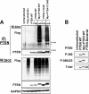
Cellular assay for PTEN ubiquitination. A, PTEN-null U87MG glioblastoma cells were transfected with expression vectors encoding FLAG-ubiquitin (Flag-Ub) either alone or in combination with a vector encoding untagged wild-type PTEN (PTEN WT). After 24 h, a sample of cells coexpressing FLAG-ubiquitin and PTEN was then treated with 10 μm PSI for 2 h. Cells were lysed, and PTEN was immunoprecipitated (IP) before Western blotting (W.blot) these immunoprecipitates with antibodies raised against the FLAG epitope and PTEN. An anti-FLAG Western blot of the lysates shows cellular protein ubiquitination levels in the FLAG-ubiquitin-expressing cells. B, U87MG cells transfected as indicated with vectors encoding wild-type PTEN were treated for 24 h with 10 μm PSI, 10 μm calpeptin, or 30 μm DMAT before PTEN expression was analyzed by Western blotting for PTEN and β-actin. DMAT inhibits CK2 (22), whereas PSI inhibits the proteasome (23).
FIGURE 2.
FIGURE 2.
Mutation of the PTEN C-terminal phosphorylation sites reduces PTEN stability and enhances ubiquitination. A, the cellular stability of the wild-type PTEN (PTEN WT) and PTEN A3 (S380A/T382A/T383A) proteins was analyzed by metabolic 35S-amino acid labeling of PTEN-null U87MG cells transduced with viruses encoding either wild-type PTEN or PTEN A3 and chasing with unlabeled amino acid-containing growth medium for the indicated times. PTEN proteins were visualized by autoradiography after immunoprecipitation. B, U87MG cells transiently expressing PTEN A3 were treated as indicated for 24 h with either 10 μm PSI or 10 μm calpeptin before Western blotting for PTEN expression along with a β-actin control. C, the ubiquitination of wild-type PTEN and PTEN A3 was compared in U87MG cells. U87MG cells were transfected with vectors encoding FLAG-tagged ubiquitin and either wild-type PTEN or PTEN A3 24 h before cells were treated with PSI at 10 μm for 2 h. Cells were lysed, and PTEN was immunoprecipitated (IP) before Western blotting (W.blot) these immunoprecipitates and the lysates with antibodies raised against the FLAG epitope, PTEN, and β-actin.
FIGURE 3.
FIGURE 3.
Membrane targeting through N-terminal myristoylation enhances PTEN ubiquitination. A, U87MG cells were transfected with vectors encoding FLAG-tagged ubiquitin (Flag-ub) and either untagged wild-type PTEN (PTEN WT) or N-terminally myristoylated wild-type (Myr-PTEN WT) or phosphatase-dead (Myr-PTEN C124S) PTEN. An N-terminal myristoylation signal and linker (33 amino acids in total) was used to target these fusion proteins to the plasma membrane, resulting in a larger slower migrating protein. Cells were lysed, and PTEN was immunoprecipitated (IP) before Western blotting (W.blot) these immunoprecipitates with antibodies raised against the FLAG epitope and PTEN. An anti-FLAG Western blot of the lysates shows cellular ubiquitination levels in the FLAG-ubiquitin-expressing cells. GAPDH, glyceraldehyde-3-phosphate dehydrogenase. B, equal amounts of immunoprecipitated wild-type PTEN and myristoylated wild-type PTEN were analyzed along with unphosphorylated bacterially expressed PTEN using phosphospecific antibodies for the C-terminal CK2 phosphorylation sites of PTEN and total protein antibodies.
FIGURE 4.
FIGURE 4.
PTEN phosphorylation does not inhibit subsequent ubiquitination in vitro. A, PTEN was phosphorylated in vitro to a stoichiometry of >3 or sham-phosphorylated in the presence or absence of CK2 enzyme as described previously (28). These proteins were then analyzed by Western blotting (W.blot) with anti-PTEN and anti-phospho-Ser380/Thr382/Thr383 PTEN antibodies. B and C, phosphorylated and sham-phosphorylated PTEN proteins were immunoprecipitated and subjected to a ubiquitination assay in vitro using either purified ubiquitin (B) or the ubiquitin K all R mutant (C). Ubiquitination of PTEN was investigated by Western blotting for PTEN.
FIGURE 5.
FIGURE 5.
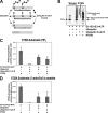
PTEN ubiquitination in vitro inhibits phosphatase activity. A, a scheme is shown that distinguishes among polyubiquitination, multiubiquitination (multisite monoubiquitination), and monoubiquitination of different lysine (K) residues on a ubiquitinated substrate protein. Using a ubiquitin mutant with each lysine residue mutated to arginine (ubiquitin K all R mutant), the formation of polyubiquitin chains is blocked. B, an anti-PTEN Western blot (W.blot) is shown comparing an in vitro ubiquitinated purified PTEN protein sample with a similar reaction using the ubiquitin K all R mutant and with a sham-ubiquitinated sample. Other immunoreactive bands within this negative control sample represent residual GST-tagged PTEN after an incomplete cleavage of this affinity purification tag. C and D, PTEN was immunoprecipitated from these ubiquitination and sham ubiquitination reactions along with a no-PTEN control, and each immunoprecipitate was assayed in vitro against PtdInsP3 (PIP3) vesicles (C) or the peptide substrate poly(Glu-Tyr(P)) (D). The mean phosphatase activity ± S.D. from triplicate samples is shown. Asterisks represent statistically significant reductions in activity compared with the relevant sham-ubiquitinated control (p < 0.05). In control blots, ubiquitination did not interfere with the efficiency of the immunoprecipitation (supplemental Fig. S2).
FIGURE 6.
FIGURE 6.
Osmotic stress induces PTEN ubiquitination and increases PtdInsP3 levels. HEK293T cells were transfected as indicated with vectors encoding wild-type PTEN (PTEN WT) and/or FLAG-tagged ubiquitin (Flag-Ub). A, cells were treated with 0.5 m sorbitol for 1, 2, or 4 h. Cells were then lysed, and PTEN was immunoprecipitated (IP) before Western blotting (W.blot) these immunoprecipitates and lysates with antibodies raised against the FLAG epitope, PTEN, and glyceraldehyde-3-phosphate dehydrogenase (GAPDH). B, cells were treated with sorbitol (0.5 m, 4 h) alone or in the presence of PSI (10 μm), and PTEN expression was analyzed by Western blotting along with a glyceraldehyde-3-phosphate dehydrogenase loading control. C, cells were treated with sorbitol as indicated (0.5 m, 10 min to 4 h). Cellular PtdInsP3 (PIP3) content was measured using a time-resolved fluorescence resonance energy transfer sensor complex by comparison with a standard curve of known PtdInsP3 concentrations as described previously (41). Parallel lysates were analyzed for the phosphorylation of Akt, PTEN, and the hyperosmotically responsive protein kinase p38 MAPK by Western blotting with phosphospecific and total protein antibodies. In these experiments, ∼60% of cells were successfully transfected. D, after transfection as indicated, some cells were treated with 0.5 m sorbitol for 1 h as indicated, and two sets of samples were then washed with normal medium lacking sorbitol for 10 or 30 min. Cells were lysed, and PTEN was immunoprecipitated before Western blotting of these immunoprecipitates and lysates with antibodies raised against the FLAG epitope, PTEN, and glyceraldehyde-3-phosphate dehydrogenase. E, cells transfected as indicated with wild-type PTEN were treated with sorbitol for 1 h before some samples were washed with normal medium for 10 or 30 min. Cellular PtdInsP3 content was then assessed as described previously (41).

Similar articles

Cited by

References

    1. Carracedo A., Pandolfi P. P. (2008) Oncogene 27, 5527–5541 - PubMed
    1. Maehama T., Dixon J. E. (1998) J. Biol. Chem. 273, 13375–13378 - PubMed
    1. Keniry M., Parsons R. (2008) Oncogene 27, 5477–5485 - PubMed
    1. Kwabi-Addo B., Giri D., Schmidt K., Podsypanina K., Parsons R., Greenberg N., Ittmann M. (2001) Proc. Natl. Acad. Sci. U.S.A. 98, 11563–11568 - PMC - PubMed
    1. Salmena L., Carracedo A., Pandolfi P. P. (2008) Cell 133, 403–414 - PubMed

Publication types

MeSH terms

Substances