

Brain-water diffusion coefficients reflect the severity of inherited prion disease
- PMID: 20177119
- PMCID: PMC2830920
- DOI: 10.1212/WNL.0b013e3181d0cc47
Brain-water diffusion coefficients reflect the severity of inherited prion disease
Abstract
Objective: Inherited prion diseases are progressive neurodegenerative conditions, characterized by cerebral spongiosis, gliosis, and neuronal loss, caused by mutations within the prion protein (PRNP) gene. We wished to assess the potential of diffusion-weighted MRI as a biomarker of disease severity in inherited prion diseases.
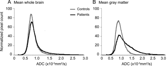
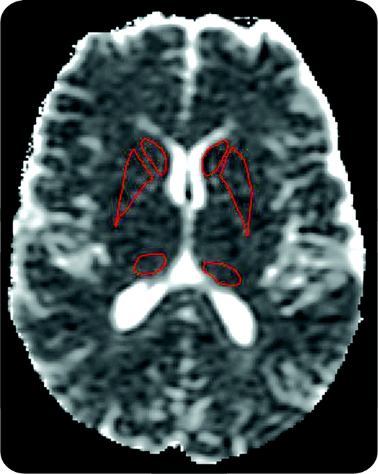
Methods: Twenty-five subjects (mean age 45.2 years) with a known PRNP mutation including 19 symptomatic patients, 6 gene-positive asymptomatic subjects, and 7 controls (mean age 54.1 years) underwent conventional and diffusion-weighted MRI. An index of normalized brain volume (NBV) and region of interest (ROI) mean apparent diffusion coefficient (ADC) for the head of caudate, putamen, and pulvinar nuclei were recorded. ADC histograms were computed for whole brain (WB) and gray matter (GM) tissue fractions. Clinical assessment utilized standardized clinical scores. Mann-Whitney U test and regression analyses were performed.
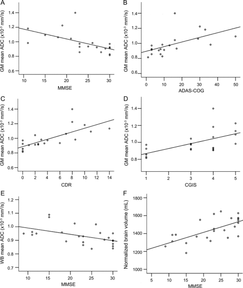
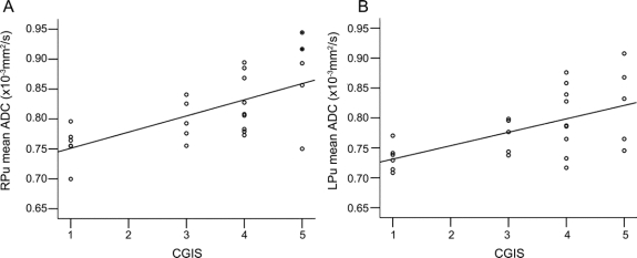
Results: Symptomatic patients exhibited an increased WB mean ADC (p = 0.006) and GM mean ADC (p = 0.024) compared to controls. Decreased NBV and increased mean ADC measures significantly correlated with clinical measures of disease severity. Using a stepwise multivariate regression procedure, GM mean ADC was an independent predictor of Clinician's Dementia Rating score (p = 0.001), Barthel Index of activities of daily living (p = 0.001), and Rankin disability score (p = 0.019).
Conclusions: Brain volume loss in inherited prion diseases is accompanied by increased cerebral apparent diffusion coefficient (ADC), correlating with increased disease severity. The association between gray matter ADC and clinical neurologic status suggests this measure may prove a useful biomarker of disease activity in inherited prion diseases.
Figures
Similar articles
-
Mathematical models for the diffusion magnetic resonance signal abnormality in patients with prion diseases.Neuroimage Clin. 2014 Nov 29;7:142-54. doi: 10.1016/j.nicl.2014.11.017. eCollection 2015. Neuroimage Clin. 2014. PMID: 25610776 Free PMC article.
-
Magnetization transfer ratio may be a surrogate of spongiform change in human prion diseases.Brain. 2010 Oct;133(10):3058-68. doi: 10.1093/brain/awq243. Brain. 2010. PMID: 20881162
-
Thalamo-striatal diffusion reductions precede disease onset in prion mutation carriers.Brain. 2009 Oct;132(Pt 10):2680-7. doi: 10.1093/brain/awp064. Epub 2009 Mar 24. Brain. 2009. PMID: 19321460 Free PMC article.
-
Diffusion-weighted MR of the brain: methodology and clinical application.Radiol Med. 2005 Mar;109(3):155-97. Radiol Med. 2005. PMID: 15775887 Review. English, Italian.
-
The genetics of prion diseases.Genet Med. 2010 Apr;12(4):187-95. doi: 10.1097/GIM.0b013e3181cd7374. Genet Med. 2010. PMID: 20216075 Review.
Cited by
-
Application of quantitative DTI metrics in sporadic CJD.Neuroimage Clin. 2014 Jan 31;4:426-35. doi: 10.1016/j.nicl.2014.01.011. eCollection 2014. Neuroimage Clin. 2014. PMID: 24624328 Free PMC article.
-
Neuroanatomical correlates of prion disease progression - a 3T longitudinal voxel-based morphometry study.Neuroimage Clin. 2016 Nov 2;13:89-96. doi: 10.1016/j.nicl.2016.10.021. eCollection 2017. Neuroimage Clin. 2016. PMID: 27942451 Free PMC article.
-
In Vivo NMR Studies of the Brain with Hereditary or Acquired Metabolic Disorders.Neurochem Res. 2015 Dec;40(12):2647-85. doi: 10.1007/s11064-015-1772-1. Epub 2015 Nov 26. Neurochem Res. 2015. PMID: 26610379 Review.
-
Mathematical models for the diffusion magnetic resonance signal abnormality in patients with prion diseases.Neuroimage Clin. 2014 Nov 29;7:142-54. doi: 10.1016/j.nicl.2014.11.017. eCollection 2015. Neuroimage Clin. 2014. PMID: 25610776 Free PMC article.
-
Multiparameter MR imaging in the 6-OPRI variant of inherited prion disease.AJNR Am J Neuroradiol. 2013 Sep;34(9):1723-30. doi: 10.3174/ajnr.A3504. Epub 2013 Mar 28. AJNR Am J Neuroradiol. 2013. PMID: 23538406 Free PMC article.
References
-
- Collinge J. Human prion diseases: aetiology and clinical features. In: Growdon JH, Rossor M, eds. The Dementias. Newton, MA: Butterworth-Heinemann; 1998:113–148.
-
- Mallucci G, Collinge J. Update on Creutzfeldt-Jakob disease. Curr Opin Neurol 2004;17:641–647. - PubMed
-
- Wroe SJ, Pal S, Siddique D, et al. Clinical and pre-mortem diagnosis of variant Creutzfeldt-Jakob disease associated with blood transfusion. Lancet 2006;368:2061–2067. - PubMed
-
- Tschampa HJ, Kallenberg K, Urbach H, et al. MRI in the diagnosis of sporadic Creutzfeldt-Jakob disease: a study on inter-observer agreement. Brain 2005;128:2026–2033. - PubMed
Publication types
MeSH terms
Substances
Grants and funding
LinkOut - more resources
Full Text Sources