. 2010 Apr 30;42(4):302-9.
doi: 10.3858/emm.2010.42.4.029.

A pertussis toxin sensitive G-protein-independent pathway is involved in serum amyloid A-induced formyl peptide receptor 2-mediated CCL2 production

Affiliations

A pertussis toxin sensitive G-protein-independent pathway is involved in serum amyloid A-induced formyl peptide receptor 2-mediated CCL2 production

Ha Young Lee et al. Exp Mol Med. .

Abstract

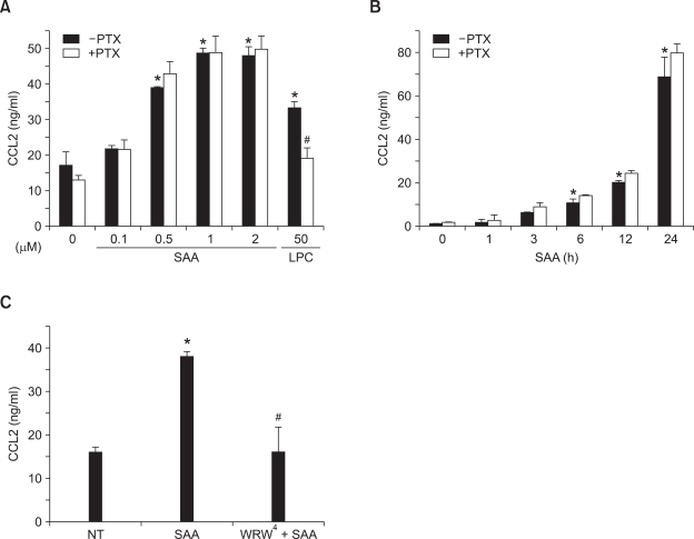
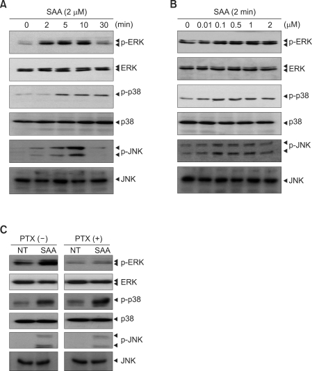
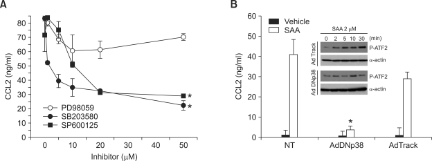
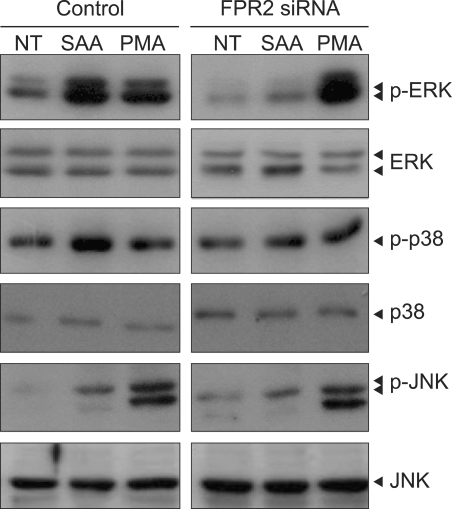
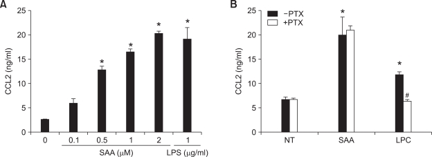
Serum amyloid A (SAA) induced CCL2 production via a pertussis toxin (PTX)-insensitive pathway in human umbilical vein endothelial cells (HUVECs). SAA induced the activation of three MAPKs (ERK, p38 MAPK, and JNK), which were completely inhibited by knock-down of formyl peptide receptor 2 (FPR2). Inhibition of p38 MAPK and JNK by their specific inhibitors (SB203580 and SP600125), or inhibition by a dominant negative mutant of p38 MAPK dramatically decreased SAA-induced CCL2 production. Inactivation of G((i)) protein(s) by PTX inhibited the activation of SAA-induced ERK, but not p38 MAPK or JNK. The results indicate that SAA stimulates FPR2-mediated activation of p38 MAPK and JNK, which are independent of a PTX-sensitive G-protein and are essential for SAA-induced CCL2 production.


Figures

Figure 1
Figure 1
SAA-induced CCL2 production from HUVECs is PTX-insensitive. HUVECs were cultured for 24 h in the presence or absence of PTX (100 ng/ml), and then stimulated for 24 h with different concentrations of SAA (0, 0.1, 0.5, 1, and 2 µM) or 50 µM of LPC (A). HUVECs were stimulated for varying times (0, 1, 3, 6, 12, and 24 h) with 2 µM SAA in the absence or presence of PTX (100 ng/ml) (B). HUVECs were stimulated for 24 h with 2 µM SAA in the absence or presence of WRW4 (10 µM) (C). The secreted CCL2 levels were determined by ELISA. The results are means ± SEM of 3 independent experiments performed in duplicate (A-C). *P < 0.05 compared to the control (0 µM). #Significantly different (P < 0.05) from treatment with LPC or SAA alone.
Figure 2
Figure 2
SAA stimulates MAPKs activities in HUVECs. (A) HUVECs were stimulated for different lengths of time (0, 2, 5, 10, and 30 min) with 2 µM of SAA. (B) HUVECs were stimulated for 5 min with different concentrations of SAA (0, 0.01, 0.1, 0.5, 1, and 2 µM). (C) HUVECs were cultured for 24 h in the presence or absence of PTX (100 ng/ml), and then stimulated for 5 min with 2 µM of SAA or vehicle. (A-C) Phosphorylated ERK, phosphorylated p38 MAPK, or phosphorylated JNK levels were determined by immunoblot analysis using anti-phospho-ERK antibody, anti-phospho-p38 MAPK antibody, or anti-phospho-JNK antibody. The results shown in (A-C) are representative of at least 3 independent experiments.
Figure 3
Figure 3
p38 MAPK and JNK are essential for SAA-induced CCL2 production by HUVECs. (A) HUVECs were pre-incubated with several concentrations (0, 1, 5, 10, 20, or 50 µM) of PD98059 (60 min), SB203580 (15 min), or SP600125 (15 min) prior to stimulation with 2 µM of SAA for 24 h. (B) The genes for GFP (AdTrack) or dominant negative p38 (AdDNp38) (MOI; 50) were transferred into HUVECs using recombinant adenoviruses prior to SAA (2 µM) stimulation for 24 h (B) or for 2, 5, 10, or 30 min (B insert). The amounts of secreted CCL2 were measured by ELISA. Phosphorylated ATF-2 levels were measured by Western blot analysis (B insert). Results are means ± SEM of 3 independent experiments performed in duplicate (A, B). *P < 0.05 compared to the control (SAA only treated).
Figure 4
Figure 4
SAA-induced MAPK activity is mediated by FPR2. HUVECs were transfected with 20 nM of FPR2 siRNA or control siRNA for 48 h. The transfected cells were stimulated with vehicle, 2 µM of SAA, or 100 nM of PMA for 5 min. Phosphorylated ERK, phosphorylated p38 MAPK, or phosphorylated JNK levels were determined by immunoblot analysis using anti-phospho-ERK antibody, anti-phospho-p38 MAPK antibody or anti-phospho-JNK antibody. The results shown are representative of 3 independent experiments.
Figure 5
Figure 5
SAA-induced CCL2 production from HUAECs is PTX-insensitive. HUAECs were stimulated for 24 h with different concentrations of SAA (0, 0.1, 0.5, 1, and 2 µM) or 1 µg/ml of LPS (A). HUAECs were cultured for 24 h in the presence or absence of PTX (100 ng/ml), and then stimulated for 24 h with 2 µM SAA or 50 µM LPC (B). The secreted CCL2 levels were determined by ELISA. Results are the means ± SEM of 3 independent experiments performed in duplicate (A, B). *P < 0.05 compared to the control (0 µM). #Significantly different (P < 0.05) from treatment with LPC alone.

Similar articles

Cited by

References

    1. Badolato R, Wang JM, Murphy WJ, Lloyd AR, Michiel DF, Bausserman LL, Kelvin DJ, Oppenheim JJ. Serum amyloid A is a chemoattractant: induction of migration, adhesion, and tissue infiltration of monocytes and polymorphonuclear leukocytes. J Exp Med. 1994;180:203–209. - PMC - PubMed
    1. Badolato R, Johnston JA, Wang JM, McVicar D, Xu LL, Oppenheim JJ, Kelvin DJ. Serum amyloid A induces calcium mobilization and chemotaxis of human monocytes by activating a pertussis toxin-sensitive signaling pathway. J Immunol. 1995;155:4004–4010. - PubMed
    1. Baranova IN, Vishnyakova TG, Bocharov AV, Kurlander R, Chen Z, Kimelman ML, Remaley AT, Csako G, Thomas F, Eggerman TL, Patterson AP. Serum amyloid A binding to CLA-1 (CD36 and LIMPII analogous-1) mediates serum amyloid A protein-induced activation of ERK1/2 and p38 mitogen-activated protein kinases. J Biol Chem. 2005;280:8031–8040. - PubMed
    1. Björkman L, Karlsson J, Karlsson A, Rabiet MJ, Boulay F, Fu H, Bylund J, Dahlgren C. Serum amyloid A mediates human neutrophil production of reactive oxygen species through a receptor independent of formyl peptide receptor like-1. J Leukoc Biol. 2008;83:245–253. - PubMed
    1. Cai L, de Beer MC, de Beer FC, van der Westhuyzen DR. Serum amyloid A is a ligand for scavenger receptor class B type I and inhibits high density lipoprotein binding and selective lipid uptake. J Biol Chem. 2005;280:2954–2961. - PubMed

Publication types

MeSH terms

LinkOut - more resources