. 2010 May;88(5):467-75.
doi: 10.1007/s00109-010-0596-3. Epub 2010 Feb 23.

Dietary magnesium, not calcium, prevents vascular calcification in a mouse model for pseudoxanthoma elasticum

Affiliations

Dietary magnesium, not calcium, prevents vascular calcification in a mouse model for pseudoxanthoma elasticum

Theo G M F Gorgels et al. J Mol Med (Berl). 2010 May.

Abstract

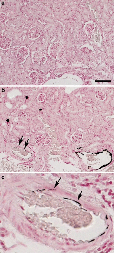
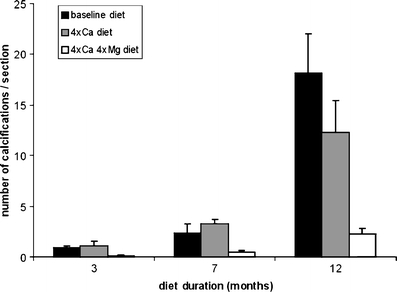
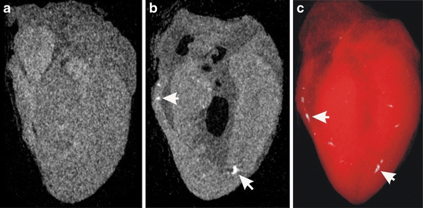
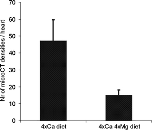
Pseudoxanthoma elasticum (PXE) is a heritable disorder characterized by ectopic calcification of connective tissue in skin, Bruch's membrane of the eye, and walls of blood vessels. PXE is caused by mutations in the ABCC6 gene, but the exact etiology is still unknown. While observations on patients suggest that high calcium intake worsens the clinical symptoms, the patient organization PXE International has published the dietary advice to increase calcium intake in combination with increased magnesium intake. To obtain more data on this controversial issue, we examined the effect of dietary calcium and magnesium in the Abcc6(-/-) mouse, a PXE mouse model which mimics the clinical features of PXE. Abcc6(-/-) mice were placed on specific diets for 3, 7, and 12 months. Disease severity was measured by quantifying calcification of blood vessels in the kidney. Raising the calcium content in the diet from 0.5% to 2% did not change disease severity. In contrast, simultaneous increase of both calcium (from 0.5% to 2.0%) and magnesium (from 0.05% to 0.2%) slowed down the calcification significantly. Our present findings that increase in dietary magnesium reduces vascular calcification in a mouse model for PXE should stimulate further studies to establish a dietary intervention for PXE.


Figures

Fig. 1
Fig. 1
Light micrographs of sections of the kidney cortex of a wild-type (a) and an Abcc6 −/− mouse (b, with detail in c), stained for calcification with von Kossa. The mice were killed after 12 months of a diet supplemented with Ca (4×Ca diet). Extensive calcification (black deposits) is present in arteries in the kidney cortex of the Abcc6 −/− mouse (b). Please notice that the calcification is located within the wall of the artery (arrows in b and c, which shows a detail of b). Bar represents 100 μm
Fig. 2
Fig. 2
Effect of diet on the number of calcifications in blood vessels in the kidney cortex of Abcc6 −/− mice. Histograms represent the average number of calcifications per kidney section as function of diet and diet duration. At each diet duration, the diet supplemented with Ca and Mg (4×Ca, 4×Mg diet) caused less calcifications than the other diets (Kruskal–Wallis test, P < 0.05)
Fig. 3
Fig. 3
Calcification in the heart analyzed by μCT. a, b Reconstructed μCT cross-section of the heart of a wild-type (a) and an Abcc6 −/− mouse (b) killed after 12 months of 4×Ca diet. Please notice in the Abcc6 −/− mouse the white dots (arrows) representing the X-ray opaque calcifications in the ventricle wall. c Three-dimensional reconstruction of the μCT scan of this Abcc6 −/− mouse, showing the distribution of the calcifications (white dots) in the heart
Fig. 4
Fig. 4
a Micrographs of paraffin sections of the ventricle of the heart of a wild-type (a) and an Abcc6 −/− mouse (b). Von Kossa staining demonstrates calcifications (black deposits; arrows) in walls of arteries in the Abcc6 −/− mouse. Bar is 50 μm
Fig. 5
Fig. 5
Histogram of the number of calcifications (±SD) in the heart, determined by μCT, as function of the diet. The 4×Ca diet caused more calcifications than the 4×Ca, 4×Mg diet (t test, P < 0.05). Diet duration was 12 months

Comment in

Similar articles

Cited by

References

    1. Hu X, Plomp AS, van Soest S, Wijnholds J, de Jong PT, Bergen AA. Pseudoxanthoma elasticum: a clinical, histopathological, and molecular update. Surv Ophthalmol. 2003;48:424–438. doi: 10.1016/S0039-6257(03)00053-5. - DOI - PubMed
    1. Finger RP, Charbel IP, Ladewig MS, Gotting C, Szliska C, Scholl HP, Holz FG. Pseudoxanthoma elasticum: genetics, clinical manifestations and therapeutic approaches. Surv Ophthalmol. 2009;54:272–285. doi: 10.1016/j.survophthal.2008.12.006. - DOI - PubMed
    1. Gheduzzi D, Sammarco R, Quaglino D, Bercovitch L, Terry S, Taylor W, Ronchetti IP. Extracutaneous ultrastructural alterations in pseudoxanthoma elasticum. Ultrastruct Pathol. 2003;27:375–384. - PubMed
    1. Chassaing N, Martin L, Calvas P, Le Bert M, Hovnanian A (2005) Pseudoxanthoma elasticum: a clinical, pathophysiological and genetic update including 11 novel ABCC6 mutations. J Med Genet 42:881–892 - PMC - PubMed
    1. Plomp AS, Hu X, de Jong PT, Bergen AA. Does autosomal dominant pseudoxanthoma elasticum exist? Am J Med Genet. 2004;126A:403–412. doi: 10.1002/ajmg.a.20632. - DOI - PubMed

Publication types

MeSH terms

LinkOut - more resources