Comparative Study
. 2010 Apr;27(4):131-40.
doi: 10.1007/s10815-010-9394-7. Epub 2010 Feb 23.

Qualitative and morphometric analysis of the ultrastructure of human oocytes cryopreserved by two alternative slow cooling protocols

Affiliations
Comparative Study

Qualitative and morphometric analysis of the ultrastructure of human oocytes cryopreserved by two alternative slow cooling protocols

Giovanni Coticchio et al. J Assist Reprod Genet. 2010 Apr.

Abstract

Purpose: To ascertain possible cell damage from cryopreservation, the ultrastructure of human oocytes cryopreserved by slow cooling was assessed.

Materials and methods: Cryopreservation was performed through two protocols with one-step or two-step propanediol. Fresh control oocytes were examined for comparison. Samples were processed for transmission electron microscopy analysis.

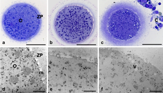
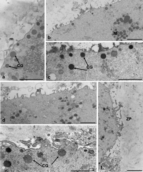
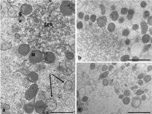
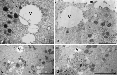
Results: By light microscopy, both fresh and frozen-thawed oocytes appeared regularly rounded, with intact zona pellucida, and homogeneous cytoplasm. By electron microscopy observation, organelles were abundant and uniformly dispersed. Mitochondria-smooth endoplasmic reticulum associations appeared regular. However, both the amount and density of cortical granules appeared abnormally reduced in frozen-thawed samples. Slight to moderate vacuolization was also found in the ooplasm of oocytes of both frozen groups.

Conclusions: Slow cooling ensures a good overall preservation of human oocytes. However, cytoplasmic vacuolization and cortical granule loss appears associated with cryopreservation, irrespective of the protocol used.


Figures

Fig. 1
Fig. 1
Fresh control oocytes (a,d); cryopreserved, one-step PrOH dehydrated oocytes (b,e); cryopreserved, two-step PrOH dehydrated oocytes (c,f). The general morphology and organelle microtopography are shown by light microscopy (LM) (ac) and transmission electron microscopy (TEM) (df). No overt difference in shape, dimensions and organelle distribution was observed among fresh and cryopreserved oocytes. The apparent reduced dimensions and increased zona pellucida thickness of the oocyte shown in c is an effect of the section plane (not equatorial). Clear vacuoles were detected by LM in cryopreserved oocytes, located in both inner (b) and outer, peripheral (c) areas. These vacuoles (V), when observed by TEM, appear as empty, scarcely electrondense structures (f). a, O: oocyte; ZP: zona pellucida. c, C: residual corona cells. d, O: oocyte; ZP: zona pellucida; m: oocyte microvilli. Bar is: 45 μm (a); 40 μm (bc); 4 μm (d); 3 μm (e); 5 μm (f)
Fig. 2
Fig. 2
Fresh control oocyte (a); cryopreserved, one-step PrOH dehydrated oocytes (b,c); cryopreserved, two-step PrOH dehydrated oocytes (df). A rim of electrondense cortical granules (CG plus arrows) is seen just beneath the oolemma in a (fresh oocyte). Cortical granules form a discontinuous layer in cryopreserved oocytes, being either grouped in some areas (CG plus arrows, c,e) or almost totally absent in other (b,d). Areas of increased density are present in the inner aspect of the zona pellucida (ZP) in f. The dense ZP corresponds to an area of the cortical ooplasm virtually devoid of cortical granules. a,d,e, m: oocyte microvilli. Bar is: 1.4 μm (a); 1.8 μm (b,d); 1 μm (c); 0.8 μm (e); 2 μm (f)
Fig. 3
Fig. 3
Fresh control oocyte (a); cryopreserved, one-step PrOH dehydrated oocyte (b); cryopreserved, two-step PrOH dehydrated oocyte (c). Mitochondria (M, a) are generally rounded and with few peripheral or transverse cristae. Voluminous aggregates between mitochondria and elements of smooth endoplasmic reticulum (SER, a) are seen in fresh and cryopreserved samples. Small cisternae presumably deriving from dilated smooth endoplasmic reticulum (arrows, a) are also commonly observed. Bar is: 0.8 μm (a); 1 μm (b); 1.2 μm (c)
Fig. 4
Fig. 4
Cryopreserved, one-step PrOH dehydrated oocytes (a,b); cryopreserved, two-step PrOH dehydrated oocytes (c,d). Numerous clear vacuoles (V) varying in dimensions are seen in the ooplasm. The vacuoles appear empty, with a limited, and often interrupted, membrane. A secondary lysosome (Ly) is observed in association with vacuoles in d. b, m: microvilli. Bar is: 1 μm (a); 1.4 μm (b); 2 μm (c,d)

Similar articles

Cited by

References

    1. Chen C. Pregnancy after human oocyte cryopreservation. Lancet. 1986;1:884–886. doi: 10.1016/S0140-6736(86)90989-X. - DOI - PubMed
    1. Al-Hasani S, Diedrich K, Ven H, Reinecke A, Hartje M, Krebs D. Cryopreservation of human oocytes. Hum Reprod. 1987;2:695–700. - PubMed
    1. Bianchi V, Coticchio G, Distratis V, Giusto N, Flamigni C, Borini A. Differential sucrose concentration during dehydration (0.2 mol/l) and rehydration (0.3 mol/l) increases the implantation rate of frozen human oocytes. Reprod Biomed Online. 2007;14:64–71. - PubMed
    1. Fabbri R, Porcu E, Marsella T, Rocchetta G, Venturoli S, Flamigni C. Human oocyte cryopreservation: new perspectives regarding oocyte survival. Hum Reprod. 2001;16:411–416. doi: 10.1093/humrep/16.3.411. - DOI - PubMed
    1. Stachecki JJ, Cohen J, Garrisi J, Munne S, Burgess C, Willadsen SM. Cryopreservation of unfertilized human oocytes. Reprod Biomed Online. 2006;13:222–227. - PubMed

Publication types

LinkOut - more resources