. 2010 May;62(5):1338-48.
doi: 10.1002/art.27410.

Induction of CD44 cleavage in articular chondrocytes

Affiliations

Induction of CD44 cleavage in articular chondrocytes

Nobunori Takahashi et al. Arthritis Rheum. 2010 May.

Abstract

Objective: The hyaluronan receptor CD44 provides chondrocytes with a mechanism for sensing and responding to changes in the extracellular matrix. The purpose of this study was to document the fragmentation and loss of CD44 and to determine the likely mechanisms involved.

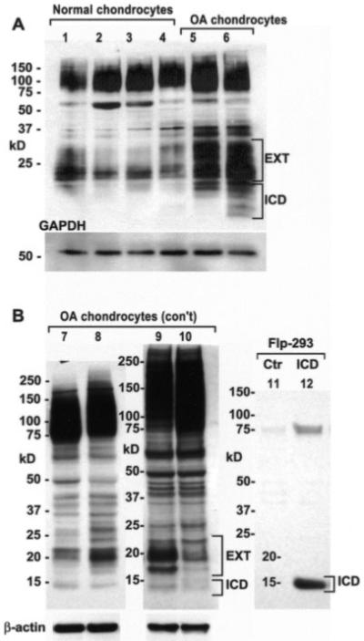
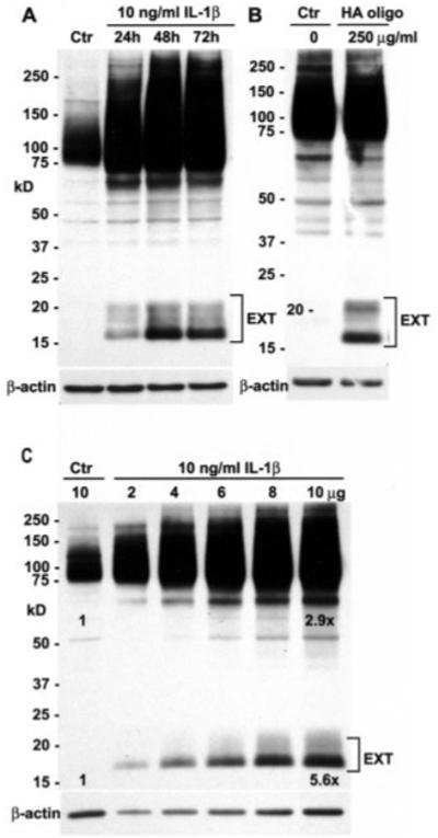
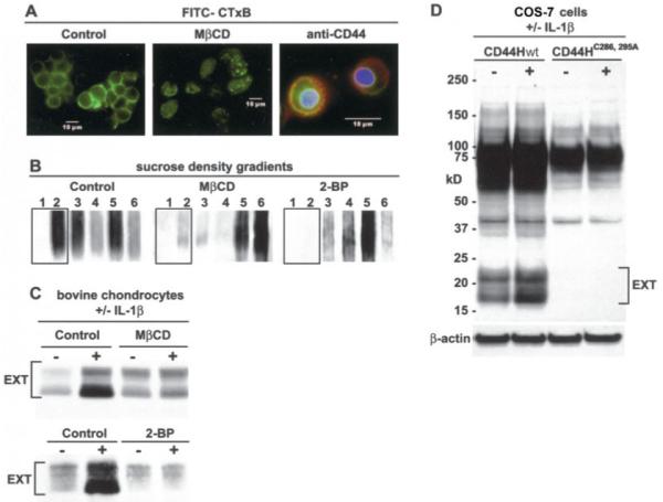
Methods: A polyclonal anti-CD44 cytotail antibody was generated to detect CD44 fragmentation by Western blot analysis. Chondrocytes were isolated from human or bovine articular cartilage. Primary articular chondrocytes were treated with interleukin-1beta (IL-1beta), hyaluronan oligosaccharides, or phorbol myristate acetate or were passaged and subcultured in monolayer to induce dedifferentiation. Conditions that altered the capacity of CD44 to transit into lipid rafts, or pharmacologic inhibitors of metalloproteinase or gamma-secretase activity were used to define the mechanism of fragmentation of CD44.

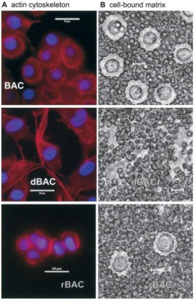
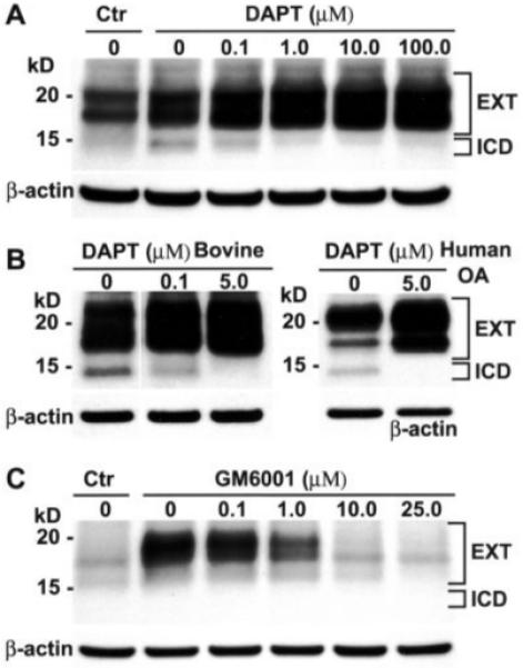
Results: Chondrocytes from osteoarthritic cartilage exhibited CD44 fragmentation as low molecular mass bands, corresponding to the CD44-EXT and CD44-ICD bands. Following dedifferentiation of chondrocytes or treatment of primary chondrocytes with hyaluronan oligosaccharides, IL-1beta, or phorbol myristate acetate, CD44 fragmentation was enhanced. Subsequent culture of the dedifferentiated chondrocytes in 3-dimensional alginate beads rescued the chondrocyte phenotype and diminished the fragmentation of CD44. Fragmentation of CD44 in chondrocytes was blocked in the presence of the metalloproteinase inhibitor GM6001 and the gamma-secretase inhibitor DAPT.

Conclusion: CD44 fragmentation, consistent with a signature pattern reported for sequential metalloproteinase/gamma-secretase cleavage of CD44, is a common metabolic feature of chondrocytes that have undergone dedifferentiation in vitro and osteoarthritic chondrocytes. Transit of CD44 into lipid rafts may be required for its fragmentation.


Figures

Figure 1
Figure 1
A, Rhodamine–phalloidin staining of permeabilized chondrocytes. Top, Primary bovine articular chondrocytes (BACs) exhibited a more rounded morphology, driven in part by cortical organization of the actin cytoskeleton. Middle, After passage in culture, dedifferentiated BACs (dBAC) became elongated and exhibited a fibroblast-like morphology, including the appearance of stress fibers. Bottom, Following culture in alginate beads for 2 weeks, redifferentiated BACs (rBAC) regained a spherical morphology, lacked stress fibers, and established a fine cortical actin network. B, Pericellular matrix on living chondrocytes, as revealed by particle exclusion assay. Top, Primary BACs synthesized and retained a pericellular matrix. Middle, With dedifferentiation, chondrocytes lost the capacity to assemble and retain a pericellular matrix. Bottom, When dedifferentiated chondrocytes were subcultured in alginate beads for 2 weeks and released from the beads, the redifferentiated chondrocytes regained a pericellular matrix. All bars = 20 μm.
Figure 2
Figure 2
Changes in gene expression and CD44 fragmentation by subculturing chondrocytes in monolayer or alginate beads. A, Dedifferentiation in culture. Total RNA from cultures of primary bovine articular chondrocytes (P0; set at 1.0) or subcultured chondrocytes from P1 to P4 were subjected to real-time reverse transcription–polymerase chain reaction (RT-PCR) analysis. B, Redifferentiation in alginate beads. Chondrocytes from passage 4 were subcultured in monolayer (R0; set at 1.0) or in alginate beads for 1 week (R1) or 2 weeks (R2). Total RNA was isolated and subjected to RT-PCR analysis. C, Western blot analysis of CD44 expression. CD44 in lysates from human knee articular chondrocytes, grown in alginate beads, was detected with BU52 (lane 1) or anti-CD44 cytotail (lane 2). CD44 in lysates from primary bovine articular chondrocytes was detected with anti-CD44 cytotail (lane 3). CD44 in lysates from primary bovine chondrocytes (P0), subcultured chondrocytes (P1–P5), alginate bead–redifferentiated chondrocytes (R1 and R2), or lysates from dedifferentiated bovine articular chondrocytes treated with control small interfering RNA (siRNA; Ctr) or CD44-specific siRNA was detected with anti-CD44 cytotail antibody. Bars represent the mean and SD fold change in mRNA copy number. HAS2 = hyaluronan synthase 2.
Figure 3
Figure 3
Western blots showing that adult human articular chondrocytes exhibit low molecular mass bands for CD44. Cell lysates were analyzed by Western blotting using anti-CD44 cytotail antisera in 3 separate experiments. A, Lysates from chondrocytes derived from ankle cartilage (grade 1 [lanes 1 and 2] and grade 2 [lane 3]) or knee cartilage (grade 1 [lane 4]) exhibited a CD44 profile with low molecular mass bands. Lysates from primary cultures of chondrocytes derived from osteoarthritic (OA) cartilage displayed a more pronounced banding pattern, with bands for both CD44-EXT and CD44-ICD (lanes 5 and 6). B, Lysates from primary cultures of chondrocytes derived from OA cartilage displayed a more pronounced banding pattern, with a CD44-ICD band at ~15 kd (lanes 7–10). The ~15-kd band observed in lysates of OA chondrocytes is equivalent in size to a recombinant human CD44-ICD (lane ICD) expressed in Flp-In–293 cells, detected using the same anti-CD44 cytotail antisera. Ctr = control.
Figure 4
Figure 4
Induction of CD44 fragmentation in bovine articular chondrocytes. Cell lysates were analyzed by Western blotting using anti-CD44 cytotail antisera. A, Upon treatment of primary bovine articular chondrocytes with 10 ng/ml interleukin-1β (IL-1β), CD44 fragmentation was enhanced, with maximal CD44-EXT formation occurring on day 2 of treatment. B, Treatment of bovine articular chondrocytes with hyaluronan oligosaccharides (HA oligo) also effected enhanced expression of CD44-EXT bands. C, Lysates (2–10 μg protein) from chondrocytes treated for 48 hours with IL-1β were analyzed on Western blots and scanned by densitometry. With values in untreated control chondrocytes (Ctr; 10 μg) set to 1.0, the full-length CD44 band in IL-1β–treated chondrocytes (after correcting for dilutions of the lysates) increased by 2.9-fold (y = 0.16x + 1.36; R2 = 0.96). After IL-1β treatment, the CD44-EXT band increased by an average of 5.6-fold (y = 0.43x + 1.30; R2 = 0.99) compared with control.
Figure 5
Figure 5
Blocking of CD44 fragmentation by inhibitors of matrix metalloproteinase and γ-secretase activities. A, CD44 fragmentation was induced by pretreatment of primary bovine articular chondrocytes either without (Ctr) or with phorbol myristate acetate (PMA). The PMA-stimulated bovine articular chondrocytes were pretreated with varying concentrations (0–100 μM) of the γ-secretase inhibitor DAPT. At a DAPT concentration of >1 μM, the 15-kd band (CD44-ICD) was no longer detectable, concomitant with an increase in the 17-20-kd doublet bands (CD44-EXT). B, In another series of experiments with PMA-stimulated primary bovine articular chondrocytes, DAPT was again partially effective at 0.1 μM but completely inhibited generation of the 15-kd band at 5.0 μM (left). In high-density cultures of human osteoarthritis (OA) chondrocytes, expression of the ~15-kd band of CD44 was blocked when the cells were treated with 5.0 μM DAPT (right). C, Primary bovine articular chondrocytes were preincubated either without (Ctr) or with 5.0 μM DAPT followed by treatment with 0–25 μM GM6001, a general matrix metalloproteinase inhibitor. Generation of the CD44-EXT bands was blocked by GM6001.
Figure 6
Figure 6
CD44 fragmentation requires transit of CD44 into lipid rafts. A, Bovine articular chondrocytes were incubated with a fluorescein isothiocyanate–labeled cholera toxin B subunit (FITC–CTxB) probe to detect lipid rafts. Preincubation with methyl-β-cyclodextrin (MβCD) eliminated surface expression of FITC–CTxB. In control chondrocytes (right panel), CD44 (red immunofluorescence) codistributed with FITC–CTxB in some regions. B, The distribution of CD44 within sucrose density gradients of lysates derived from chondrocytes pretreated with or without MβCD or 2-bromopalmitate (2-BP) was assessed by Western blotting, using anti-CD44 cytotail antisera. C, Bovine articular chondrocytes pretreated in the absence or presence of MβCD or 2-BP were stimulated with 10 ng/ml interleukin-1β (IL-1β). Both MβCD and 2-BP reduced the enhanced expression of CD44-EXT bands detected by Western blotting using anti-CD44 cytotail antisera. D, COS-7 cells were transfected with a full-length human CD44 construct (CD44Hwt) or a full-length human CD44 containing 2 cysteine–alanine mutations (CD44H-C286, 295A) followed by incubation with or without 10 ng/ml IL-1β and Western blotting using anti-CD44 cytotail antisera. Control CD44Hwt underwent cleavage, but enhanced generation of CD44-EXT followed IL-1β treatment. However, the CD44H-C286, 295A construct displayed no capacity for fragmentation in control or IL-1β–treated COS-7 transfectants.

References

    1. Hardingham TE, Fosang AJ. Proteoglycans: many forms and many functions. FASEB J. 1992;6:861–70. - PubMed
    1. Knudson W, Knudson C. An update on hyaluronan and CD44 in cartilage. Current Opin Orthop. 2004;15:369–75.
    1. Nishida Y, Knudson CB, Nietfeld JJ, Margulis A, Knudson W. Antisense inhibition of hyaluronan synthase-2 in human articular chondrocytes inhibits proteoglycan retention and matrix assembly. J Biol Chem. 1999;274:21893–9. - PubMed
    1. Knudson CB, Knudson W. Hyaluronan-binding proteins in development, tissue homeostasis and disease. FASEB J. 1993;7:1233–41. - PubMed
    1. Thorne RF, Legg JW, Isacke CM. The role of the CD44 transmembrane and cytoplasmic domains in co-ordinating adhesive and signalling events. J Cell Sci. 2004;117:373–80. - PubMed

Publication types

MeSH terms