. 2010 Apr 6;49(13):2932-42.
doi: 10.1021/bi100159p.

Characterization of the colchicine binding site on avian tubulin isotype betaVI

Affiliations

Characterization of the colchicine binding site on avian tubulin isotype betaVI

Shubhada Sharma et al. Biochemistry. .

Abstract

Tubulin, the basic component of microtubules, is present in most eukaryotic cells as multiple gene products, called isotypes. The major tubulin isotypes are highly conserved in terms of structure and drug binding capabilities. Tubulin isotype betaVI, however, is significantly divergent from the other isotypes in sequence, assembly properties, and function. It is the major beta-tubulin isotype of hematopoietic tissue and forms the microtubules of platelet marginal bands. The interaction of the major tubulin isotypes betaI, betaII, betaIII, and betaIotaV with antimicrotubule drugs has been widely studied, but little is known about the drug binding properties of tubulin isotype betaVI. In this investigation, we characterize the activity of various colchicine site ligands with tubulin isolated from Gallus gallus erythrocytes (CeTb), which is approximately 95% betaVI. Colchicine binding is thought to be a universal property of higher eukaryotic tubulin; however, we were unable to detect colchicine binding to CeTb under any experimental conditions. Podophyllotoxin and nocodazole, other colchicine site ligands with divergent structures, were able to inhibit paclitaxel-induced CeTb assembly. Surprisingly, the colchicine isomer allocolchicine also inhibited CeTb assembly and displayed measurable, moderate affinity for CeTb (K(a) = 0.18 x 10(5) M(-1) vs 5.0 x 10(5) M(-1) for bovine brain tubulin). Since allocolchicine and colchicine differ in their C ring structures, the two C ring colchicine analogues were also tested for CeTb binding. Kinetic experiments indicate that thiocolchicine and chlorocolchicine bind to CeTb, but very slowly and with low affinity. Molecular modeling of CeTb identified five divergent amino acid residues within 6 A of the colchicine binding site compared to betaI, betaII, and betaIV; three of these amino acids are also altered in betaIII-tubulin. Interestingly, the altered amino acids are in the vicinity of the A ring region of the colchicine binding site rather than the C ring region. We propose that the amino acid differences in the binding site constrict the A ring binding domain in CeTb, which interferes with the positioning of the trimethoxyphenyl A ring and prevents C ring binding site interactions from efficiently occurring. Allocolchicine is able to accommodate the altered binding mode because of its smaller ring size and more flexible C ring substituents. The sequence of the colchicine binding domain of CeTb isotype betaVI is almost identical to that of its human hematopoietic counterpart. Thus, through analysis of the interactions of ligands with CeTb, it may be possible to discover colchicine site ligands that specifically target tubulin in human hematopoietic cells.


Figures

FIGURE 1
FIGURE 1
Structures of colchicine, chlorocolchicine, thiocolchicine, podophyllotoxin, nocodazole and allocolchicine.
FIGURE 2
FIGURE 2
Sequence alignment of β subunit of 1SA0 and Gallus gallus isotype βVI. Residues colored in red are those which were within 6Å of the binding site. Residues highlighted in cyan are those in the colchicine binding domain. Mutations are marked with a star underneath the alignment.
FIGURE 3
FIGURE 3
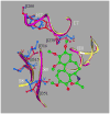
Schematic diagram of differences in mammalian brain β tubulin (from 1SA0 structure) and isotype βVI of chicken in the colchicine binding site. The 1SA0-derived coordinates of α/β heterodimer including GTP, GDP and colchicine (SH group was replaced by H) were used as starting coordinates for minimization procedure for the mammalian brain tubulin dimer. The chicken erythrocyte tubulin dimer was created from 1SA0 and the two dimers were minimized and superimposed as described under Materials and Methods. The residues containing any atom within 6Å of bound colchicine were selected. Only the divergent residues within this cut-off are shown in small ball-and-stick representation in blue (chicken isotype βVI) and brown (mammalian brain β–tubulin). The helices, β–sheets and loops for mammalian brain β–tubulin and chicken isotype βVI are shown as purple and yellow ribbons respectively. The colchicine is shown in large ball-and-stick representation with C, O and N colored green, red and dark blue, respectively. Only the mammalian structure colchicine is shown as it differs only slightly from the colchicine in the chicken βVI structure. Five substitutions; Y200-F, C239-S, A315-C, V316-I and T351-V, were observed in chicken isotype βVI relative to mammalian brain β–tubulin within the 6 Å cut-off.
FIGURE 4
FIGURE 4
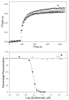
(A) Effect of colchicine on paclitaxel induced polymerization of CeTb. CeTb (5 μM) in PME buffer and 0.1 mM GTP was incubated with increasing concentrations of colchicine for 90 min at 25° C. The samples were then treated with paclitaxel (5 μM) at 37°C and the polymerization was monitored in terms of apparent absorption at 400 nm. The colchicine concentrations were 0 μM (▲), 80 μM (●), 100 μM (○), 120 μM (■) and 140 μM (□). (B) Plot for IC50 of colchicine for BbTb (3) and Cetb (3). The IC50 of BbTb was 2.7 μM and that for CeTb was >140 μM.
FIGURE 5
FIGURE 5
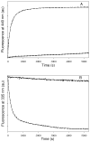
(A) Effect of BbTb (- - -) and CeTb (·····) on fluorescence of colchicine. Tubulin (1 μM) was added to colchicine (150 μM) in PME buffer in presence of 0.1 mM GTP at 25°C and enhancement in colchicine fluorescence at 440 nm was monitored as function of time. (B) Effect of colchicine on intrinsic fluorescence of BbTb (- - -) and CeTb (·····). Colchicine (150 μM) was added to tubulin (1 μM) in PME buffer in presence of 0.1 mM GTP at 25°C and the quenching of intrinsic protein fluorescence at 335 nm was monitored as function of time.
FIGURE 6
FIGURE 6
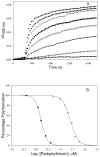
(A) Effect of podophyllotoxin on paclitaxel induced polymerization of CeTb. CeTb (5 μM) in PME buffer and 0.1 mM GTP was incubated with increasing concentrations of podophyllotoxin for 25 min at 25° C. The samples were then treated with paclitaxel (5 μM) at 37 oC and the polymerization was monitored in terms of apparent absorption at 400 nm. The podophyllotoxin concentrations were 0 μM(▲), 5 μM (●), 10 μM (○),15 μM (■), 20 μM (□), 30 μM (★), 40 μM (☆), and 60 μM(▼;). (B) Plot for IC50 of podophyllotoxin for BbTb (●) and Cetb (○). The IC50 of BbTb was 1.6 μM and that for CeTb 25 μM.
FIGURE 7
FIGURE 7
Plot for IC50 of nocodazole for BbTb (●) and Cetb (○). Tubulin (5 μM) in PME buffer and 0.1 mM GTP was incubated with increasing concentrations of nocodazole for 40 min at 25° C. The samples were then treated with paclitaxel (5 μM) at 37°C and the polymerization was monitored in terms of apparent absorption at 350 nm. The IC50 of nocodazole for BbTb was 7.7 μM and that for CeTb was 87 μM.
FIGURE 8
FIGURE 8
Emission spectra of allocolchicine in the presence of BbTb (—) and CeTb (·····). Allocolchicine (150 μM) was incubated with tubulin (1 μM) at 25°C for 90 min and the ligand emission spectra were obtained by exciting the samples at 315 nm.
FIGURE 9
FIGURE 9
Enhancement of allocolchicine fluorescence upon binding to BbTb (●) and CeTb (▲) at 25°C as a function of ligand concentration. The excitation wavelength was 315 nm. Spectra were collected as described under Materials and Methods.
FIGURE 10
FIGURE 10
Enhancement of allocolchicine fluorescence upon binding of BbTb (- - -) and CeTb (·····) as function of time. Tubulin 1 μM was added to an allocolchicine solution (150 μM) at 25° C. The excitation and emission wavelengths were 330 and 400 nm, respectively. The kinetics of binding were evaluated as described under Materials and Methods.
FIGURE 11
FIGURE 11
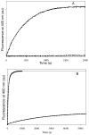
Competition of allocolchicine binding to tubulin by podophyllotoxin. The kinetics of allocolchicine binding to CeTb (A) and BbTb (B) in the absence (·····) and presence (- - -) of podophyllotoxin were measured. CeTb 1 μM was incubated with 0 μM and 150 μM podophyllotoxin for 25 min at 25° C. At the end of incubation, allocolchicine (150 μM) was added to the solution and binding was studied in terms of increase in allocolchicine fluorescence as a function of time. For experiment with BbTb, 50 μM of each ligand was used. DMSO was limited to 10%. The excitation and emission wavelengths were 330 and 400 nm, respectively.
FIGURE 12
FIGURE 12
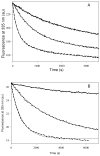
Quenching of intrinsic fluorescence of BbTb (A) and CeTb (B) by colchicine (—), thiocolchicine (·····) and chlorocolchicine (- - -), as a function of time. Ligands were added to tubulin solutions to yield a final concentration of 1 μM of tubulin and 20 μM ligands in A and 1 μM of tubulin and 150 μM ligands in B at 25°C. The excitation and emission wavelengths were 295 and 335 nm, respectively.

Similar articles

Cited by

References

    1. Morejohn LC, Fosket DE. The biochemistry of compounds with anti-microtubule activity in plant-cells. Pharmacol Ther. 1991;51:217–230. - PubMed
    1. Kohler P. The biochemical basis of anthelmintic action and resistance. Int J Parasitol. 2001;31:336–345. - PubMed
    1. Jordan A, Hadfield JA, Lawrence NJ, McGown AT. Tubulin as a target for anticancer drugs: Agents which interact with the mitotic spindle. Med Res Rev. 1998;18:259–296. - PubMed
    1. Luduena RF. Multiple forms of tubulin: Different gene products and covalent modifications, in. International Review of Cytology - a Survey of Cell Biology. 1998;178:207–275. - PubMed
    1. Verdier-Pinard P, Pasquier E, Xiao H, Burd B, Villard C, Lafitte D, Miller LM, Angeletti RH, Horwitz SB, Braguer D. Tubulin proteomics: Towards breaking the code. Anal Biochem. 2009;384:197–206. - PMC - PubMed

Publication types