. 2011 Mar;15(3):668-78.
doi: 10.1111/j.1582-4934.2010.01041.x.

Hypoxia-induced endothelial secretion of macrophage migration inhibitory factor and role in endothelial progenitor cell recruitment

Affiliations

Hypoxia-induced endothelial secretion of macrophage migration inhibitory factor and role in endothelial progenitor cell recruitment

David Simons et al. J Cell Mol Med. 2011 Mar.

Abstract

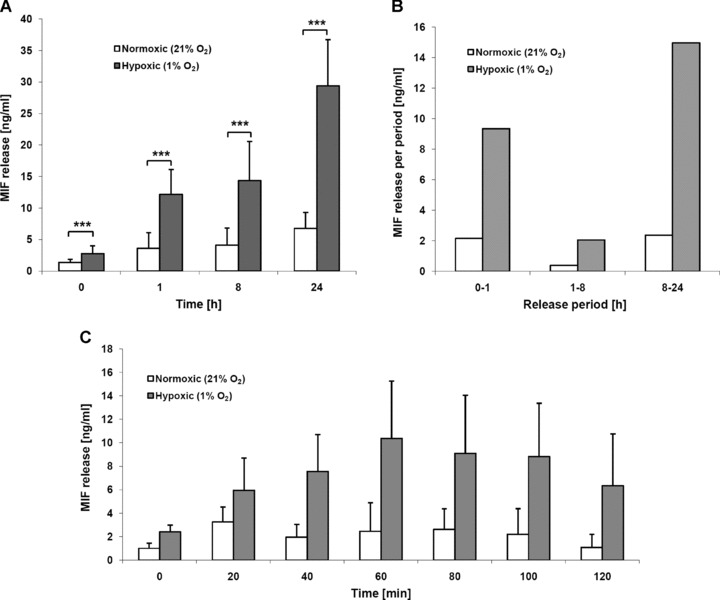
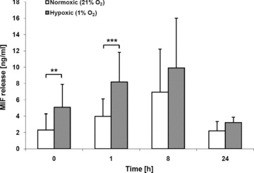
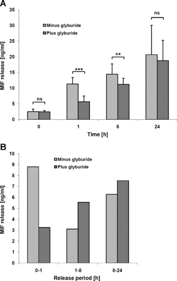
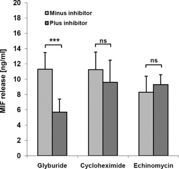
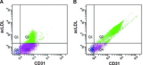
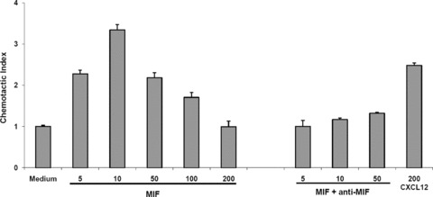
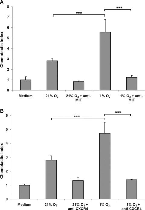
Macrophage migration inhibitory factor (MIF) is a pleiotropic inflammatory cytokine that was recently identified as a non-cognate ligand of the CXC-family chemokine receptors 2 and 4 (CXCR2 and CXCR4). MIF is expressed and secreted from endothelial cells (ECs) following atherogenic stimulation, exhibits chemokine-like properties and promotes the recruitment of leucocytes to atherogenic endothelium. CXCR4 expressed on endothelial progenitor cells (EPCs) and EC-derived CXCL12, the cognate ligand of CXCR4, have been demonstrated to be critical when EPCs are recruited to ischemic tissues. Here we studied whether hypoxic stimulation triggers MIF secretion from ECs and whether the MIF/CXCR4 axis contributes to EPC recruitment. Exposure of human umbilical vein endothelial cells (HUVECs) and human aortic endothelial cells (HAoECs) to 1% hypoxia led to the specific release of substantial amounts of MIF. Hypoxia-induced MIF release followed a biphasic behaviour. MIF secretion in the first phase peaked at 60 min. and was inhibited by glyburide, indicating that this MIF pool was secreted by a non-classical mechanism and originated from pre-formed MIF stores. Early hypoxia-triggered MIF secretion was not inhibited by cycloheximide and echinomycin, inhibitors of general and hypoxia-inducible factor (HIF)-1α-induced protein synthesis, respectively. A second phase of MIF secretion peaked around 8 hrs and was likely due to HIF-1α-induced de novo synthesis of MIF. To functionally investigate the role of hypoxia-inducible secreted MIF on the recruitment of EPCs, we subjected human AcLDL(+) KDR(+) CD31(+) EPCs to a chemotactic MIF gradient. MIF potently promoted EPC chemotaxis in a dose-dependent bell-shaped manner (peak: 10 ng/ml MIF). Importantly, EPC migration was induced by supernatants of hypoxia-conditioned HUVECs, an effect that was completely abrogated by anti-MIF- or anti-CXCR4-antibodies. Thus, hypoxia-induced MIF secretion from ECs might play an important role in the recruitment and migration of EPCs to hypoxic tissues such as after ischemia-induced myocardial damage.


Figures

Fig 1
Fig 1
Biphasic hypoxia-induced MIF secretion from ECs. (A) Cumulative MIF release from HUVEC cells under hypoxic and normoxic conditions between 0 and 24 hrs. (B) Period-specific MIF release under hypoxic and normoxic conditions (data deduced from Fig. 1A). (C) Short-term cumulative MIF release between 0 and 120 min. 15,000 cells were incubated overnight in 24-well plates in a normoxic incubator, one plate for each time-point. Half of the plates were transferred into a hypoxic incubator, the other half remained in the normoxic incubator. At t= 0, media were replaced by media that were conditioned under either normoxic or 1% hypoxia conditions (>14 hrs). Supernatants were collected at 0, 1, 8 and 24 hrs or 0, 20, 40, 60, 80, 100 and 120 min. after medium change and MIF concentrations in the cell supernatants were determined by ELISA. Bars represent mean values, error bars refer to the corresponding standard deviations (n= 9). ***, indicates statistical significance with a power of P < 0.005.
Fig 2
Fig 2
Hypoxia-induced MIF secretion from HAoECs. Experiments were performed essentially as described in the legend to Figure 1. Bars represent mean values (n= 3) ± S.D. Asterisks, * and ***, indicate statistical significance with P < 0.05 and P < 0.005, respectively.
Fig 3
Fig 3
Inhibition of hypoxia-induced rapid MIF release by the ABCA1 transporter inhibitor glyburide. HUVECs (15,000 cells) were incubated overnight in 24-well plates in a normoxic incubator. At t= 0, media were replaced by hypoxic media containing 5 μM glyburide. (A) Cumulative hypoxia-induced MIF release in the presence or absence of 5 μM glyburide at the indicated time intervals as determined by ELISA. (B) Period-specific MIF release from HUVECs under hypoxic conditions in the absence versus presence of glyburide. Data were derived from the experiment of Figure 4A. Bars represent mean values (n= 6) ± S.D. Asterisks, ** and ***, indicate statistical significance with P < 0.01 and P < 0.005, respectively.
Fig 4
Fig 4
Hypoxia-induced rapid MIF release is not affected by cycloheximide or echinomycin. HUVECs (15,000 cells) were incubated overnight in 24-well plates in a normoxic incubator. At t= 0, media were replaced by hypoxic media either containing 5 μM glyburide, 2 μg/ml cycloheximide or 10 nM echinomycin. Solvent was used as a control. MIF concentrations were determined from 1 hr supernatants by ELISA. Bars represent mean values (n= 3) ± S.D. Asterisks, ***, indicate statistical significance with P < 0.005.
Fig 5
Fig 5
EPCs take up DiI-labelled acLDL and co-express CD31 and VEGFR2. Characterization of EPCs was performed by flow cytometric analysis. (A) Cell population obtained after 2 days on fibronectin. (B) Cell population harvested after 7 days culture on fibronectin and removal of non-adhering cells.
Fig 6
Fig 6
MIF specifically induces EPC chemotaxis. Dose-dependent enhancement of EPC chemotaxis by recombinant human MIF and blockade by neutralizing anti-MIF mAbs. Chemotaxis was evaluated in Transwell chambers. The upper chamber contained 50,000 calcein-stained EPCs. The lower chamber contained varying concentrations of recombinant human MIF (ng/ml) or recombinant human CXCL12 (ng/ml). MIF and anti-MIF antibodies or recombinant human CXCL12 were added as indicated. Bars represent mean values ± S.D. (n= 5).
Fig 7
Fig 7
Stimulation of EPC chemotaxis by hypoxia-conditioned HUVEC media and blockade by anti-MIF and anti-CXCR4 antibodies. Hypoxia-conditioned HUVEC supernatants but not normoxia-conditioned control media promotes EPC chemotaxis and this effect is abolished by anti-MIF antibodies (A). (B) as in (A), but blockade by anti-CXCR4 antibodies. The hypoxia-conditioned HUVEC media were added to the lower chamber of a Transwell device. Bars represent mean values ± S.D. (n= 3). Asterisks, ***, indicates statistical significance with P < 0.005.

Similar articles

Cited by

References

    1. Bernhagen J, Calandra T, Bucala R. Regulation of the immune response by macrophage migration inhibitory factor: biological and structural features. J Mol Med. 1998;76:151–61. - PubMed
    1. Calandra T, Roger T. Macrophage migration inhibitory factor: a regulator of innate immunity. Nat Rev Immunol. 2003;3:791–800. - PMC - PubMed
    1. Morand EF, Leech M, Bernhagen J. MIF: a new cytokine link between rheumatoid arthritis and atherosclerosis. Nat Rev Drug Discov. 2006;5:399–410. - PubMed
    1. Zernecke A, Bernhagen J, Weber C. Macrophage migration inhibitory factor in cardiovascular disease. Circulation. 2008;117:1594–602. - PubMed
    1. Bernhagen J, Krohn R, Lue H, et al. MIF is a noncognate ligand of CXC chemokine receptors in inflammatory and atherogenic cell recruitment. Nat Med. 2007;13:587–96. - PubMed

Publication types

MeSH terms