. 2010 Feb 23:10:57.
doi: 10.1186/1471-2148-10-57.

Evolution of spliceosomal introns following endosymbiotic gene transfer

Affiliations

Evolution of spliceosomal introns following endosymbiotic gene transfer

Nahal Ahmadinejad et al. BMC Evol Biol. .

Abstract

Background: Spliceosomal introns are an ancient, widespread hallmark of eukaryotic genomes. Despite much research, many questions regarding the origin and evolution of spliceosomal introns remain unsolved, partly due to the difficulty of inferring ancestral gene structures. We circumvent this problem by using genes originated by endosymbiotic gene transfer, in which an intron-less structure at the time of the transfer can be assumed.

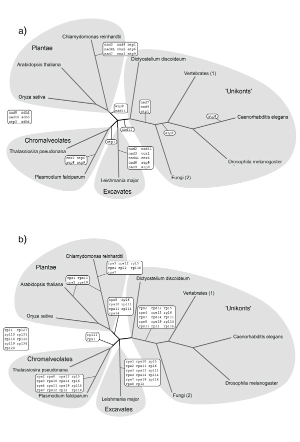
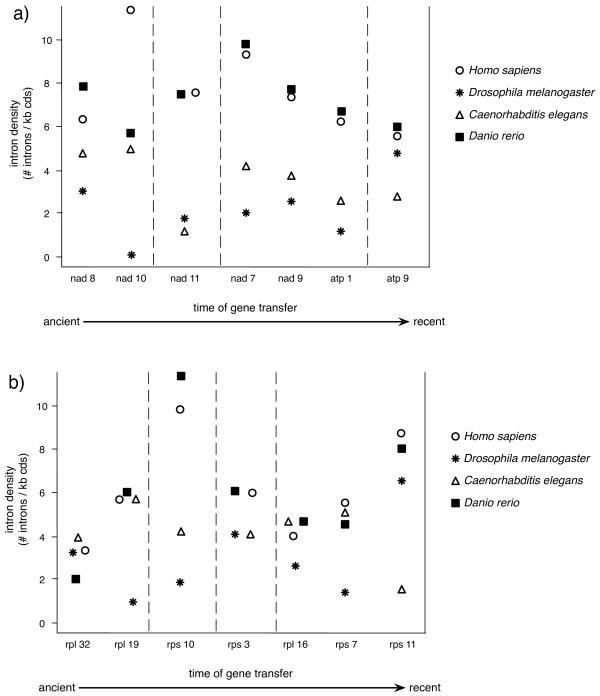
Results: By comparing the exon-intron structures of 64 mitochondrial-derived genes that were transferred to the nucleus at different evolutionary periods, we can trace the history of intron gains in different eukaryotic lineages. Our results show that the intron density of genes transferred relatively recently to the nuclear genome is similar to that of genes originated by more ancient transfers, indicating that gene structure can be rapidly shaped by intron gain after the integration of the gene into the genome and that this process is mainly determined by forces acting specifically on each lineage. We analyze 12 cases of mitochondrial-derived genes that have been transferred to the nucleus independently in more than one lineage.

Conclusions: Remarkably, the proportion of shared intron positions that were gained independently in homologous genes is similar to that proportion observed in genes that were transferred prior to the speciation event and whose shared intron positions might be due to vertical inheritance. A particular case of parallel intron gain in the nad7 gene is discussed in more detail.


Figures

Figure 1
Figure 1
Intron densities and distributions of intron phases and exon symmetry are shown for all proto-mitochondrial genes of the oxidative phosphorylation pathway and the ribosomal mitochondrial proteins and their homologs. The intron density is given as the number of introns per 1 kb of coding sequence for each species for the groups animals (cel: Caenorhabditis elegans, dme: Drosophila melanogaster, dre: Danio rerio, hsa: Homo sapiens, rno: Rattus norvegicus), fungi (afu: Aspergillus fumigatus, spo: Schizosaccharomyces pombe, sce: Saccharomyces cerevisiae, yli: Yarrowia lipolytica, cgl: Candida glabrata), protists (ddi: Dictyostelium discoideum, tps: Thalassiosira pseudonana, lma: Leishmania major, pfa: Plasmodium falciparum), and plants/green alga (cre: Chlamydomonas reinhardtii, osa: Oryza sativa, ath: Arabidopsis thaliana). The average intron densities for the different species are indicated by horizontal lines, values were taken or computed from the literature [38,57-59]. Intron phases are presented in percentages for all genes. The percentages of exon symmetry are shown separately for symmetric and asymmetric exons, in which all possible symmetries are considered.
Figure 2
Figure 2
The tree represents current view of phylogenetic relationships between the lineages sampled in this analysis, as summarized by Roger and Simpson [60]. Inferred timing for the transfers of genes from the mitochondrion to the host nucleus is labeled at the branches. The timing of each transfer depends on the presence or absence of each gene in the mitochondrial genome and the phylogeny. Proto-mitochondrial genes of a) the oxidative phosphorylation pathway, b) ribosomal mitochondrial proteins.
Figure 3
Figure 3
The intron density is shown for genes that were transferred at different time scales during evolution from the mitochondrion to the nucleus in Homo sapiens, Drosophila melanogaster, Caenorhabditis elegans and Danio rerio. a) proteins of the oxidative phosphorylation pathway, b) ribosomal mitochondrial proteins. Although the most ancient class of transfers (nad8, nad10, rpl32, rpl19) is unassigned in Figure 2 we consider them to be relatively more ancient than nad11, rps10 and rps3 because the latter are nuclear only in unikonts and the former are nuclear in most eukaryotic groups.
Figure 4
Figure 4
A comprehensive phylogeny of the gene nad7 including mitochondrial and nuclear encoded homologs. The tree is rooted by alpha-proteobacterial homologs. The two independent gene transfers of nad7 from the mitochondrion to the nucleus are labelled at the tree. The nucleotide region of the shared intron position is shown. The different lengths of the intron sequences are indicated in parentheses, the splicing sites are marked in bold

Similar articles

Cited by

References

    1. Gilbert W. Why genes in pieces? Nature. 1978;271(5645):501. doi: 10.1038/271501a0. - DOI - PubMed
    1. Nilsen TW. The spliceosome: the most complex macromolecular machine in the cell? Bioessays. 2003;25(12):1147–1149. doi: 10.1002/bies.10394. - DOI - PubMed
    1. Collins L, Penny D. Complex spliceosomal organization ancestral to extant eukaryotes. Mol Biol Evol. 2005;22(4):1053–1066. doi: 10.1093/molbev/msi091. - DOI - PubMed
    1. Gilbert W, Glynias M. On the ancient nature of introns. Gene. 1993;135(1-2):137–144. doi: 10.1016/0378-1119(93)90058-B. - DOI - PubMed
    1. Cech TR. The generality of self-splicing RNA: relationship to nuclear mRNA splicing. Cell. 1986;44(2):207–210. doi: 10.1016/0092-8674(86)90751-8. - DOI - PubMed

Publication types

LinkOut - more resources