. 2010 Feb 23:10:23.
doi: 10.1186/1471-213X-10-23.

Spatial discontinuity of optomotor-blind expression in the Drosophila wing imaginal disc disrupts epithelial architecture and promotes cell sorting

Affiliations

Spatial discontinuity of optomotor-blind expression in the Drosophila wing imaginal disc disrupts epithelial architecture and promotes cell sorting

Jie Shen et al. BMC Dev Biol. .

Abstract

Background: Decapentaplegic (Dpp) is one of the best characterized morphogens, required for dorso-ventral patterning of the Drosophila embryo and for anterior-posterior (A/P) patterning of the wing imaginal disc. In the larval wing pouch, the Dpp target gene optomotor-blind (omb) is generally assumed to be expressed in a step function above a certain threshold of Dpp signaling activity.

Results: We show that the transcription factor Omb forms, in fact, a symmetrical gradient on both sides of the A/P compartment boundary. Disruptions of the Omb gradient lead to a re-organization of the epithelial cytoskeleton and to a retraction of cells toward the basal membrane suggesting that the Omb gradient is required for correct epithelial morphology. Moreover, by analysing the shape of omb gain- and loss-of-function clones, we find that Omb promotes cell sorting along the A/P axis in a concentration-dependent manner.

Conclusions: Our findings show that Omb distribution in the wing imaginal disc is described by a gradient rather than a step function. Graded Omb expression is necessary for normal cell morphogenesis and cell affinity and sharp spatial discontinuities must be avoided to allow normal wing development.


Figures

Figure 1
Figure 1
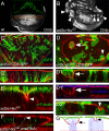
Omb expression is graded in the larval wing pouch. In this and subsequent figures, x-y scans are oriented with anterior left and dorsal up, x-z scans with apical up. (A) x-y section of wild type wing imaginal disc stained with anti-Omb. (A') Profile of fluorescence intensity along the A/P axis. (A'') Profile of fluorescence intensity along the P/D axis. (B) x-z section along the A/P axis. (C) x-z section along the P/D axis.
Figure 2
Figure 2
Cellular retraction associated with lateral tkvQD clones. (A) Wild type Omb expression pattern. The inset shows the profile of fluorescence intensity in a stripe of cells (orange box) along the A/P axis. (B) Lateral tkvQD clones (arrowheads) up-regulate Omb to a level comparable to central endogenous Omb. (C) CD8-GFP control clones, labelled by anti-GFP staining (green), show normal phalloidin staining. (C') x-z scan through the same disc. (D) Lateral tkvQD clones, labelled by CD8-GFP co-expression (green), either form a fold at the clonal border (arrow) or retract cells toward the basal side within the clone (arrow head) as revealed by phalloidin staining. (D1 and D2) x-z scans through clones marked by arrow and arrowhead, respectively, in (D). The x-z sections presented in C', D1, D1', and D2 are derived from C and D, respectively, but are shown at a 1.5-fold higher magnification. D1 shows the retraction of cells at the clonal border and D1' shows loss of the apical microtubule web in retracted cells (arrow). (E) x-z scan through tkvQD clones. The apical microtubule web, stained by anti-α-tubulin (green), is reduced in the retracting cells (arrowheads). (F) x-z scan of UAS-tkvQD UAS-ombRNAi clone. Cell shape in this lateral clone (arrowhead) which is identified by the lack of Omb appears normal. The x-y position of the clone is shown in Additional File 2B. (G) Model of cell shape changes in clones with peripherally (left) and centrally (right) retracting cells. High local microtubule density (shown in green) is found both in the peripodial membrane (squamous epithelium) and the AMW of the underlying columnar epithelium. Mutant cells are rendered with blue, wild type cells with black outlines. The left cartoon visualizes that cellular retraction at the clone boundary also leads to non-autonomous attenuation of the AMW in the flanking wild type cells. Phalloidin staining is red in all panels.
Figure 3
Figure 3
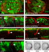
Lack of Omb causes cells to retract toward the basal side. (A) omb null mutant clones (arrowhead), labelled by absence of GFP, contain retracted cells in the clonal center. (B) ombRNAi clones (arrowhead), labelled by absence of Omb, also contain retracted cells in the clonal center. (C-G) x-z scans of omb null mutant clones (marked by absence of GFP) and ombRNAi clones (marked by reduction of Omb). (C) Central (arrowhead) but not the very lateral (arrow) mutant clones contain retracted cells. (D) ombRNAi clone (marked by reduced anti-Omb staining, arrowhead) with strong central retraction toward the basal lamina. (E and F) Reduction of the apical microtubule web in retracting cells of omb null mutant clone (arrowhead, E) and ombRNAi clone (arrowhead, F). (G) The DE-Cadherin level appears normal (arrowhead) in omb null mutant clones (absence of GFP). (H) Dorsal, middle, and ventral views of an omb mutant clone in adult wing.
Figure 4
Figure 4
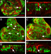
Omb overexpression causes autonomous and non-autonomous cellular retraction. Tubα1>omb clones are marked by strong anti-Omb staining (green). (A) Omb overexpressing cells tend to scatter in the epithelium. Thus, mosaic discs generally contain few cases of grouped cells (arrow). These show central retractions. (A') x-z scan through a retracting clone. (B and C) Enhanced generation of Omb overexpressing cells by more severe heat shock conditions causes clustering of wild type cells into groups with smooth outlines (arrowheads). In these non-clonal wild type cell groups, cellular retraction occurrs in the periphery (B') or in the center (C'). (A-C) are x-y scans, (A'-C') x-z scans. Arrows in A to C indicate the cell clusters that are shown in A' to C'.
Figure 5
Figure 5
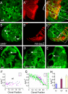
Omb regulates cell affinity in a concentration-dependent manner. Only Clones within a region corresponding to the boxed areas in A'' and B'' were selected for measurement. The position of the A/P boundary (broken line) was determined by Ci (A') or hh-lacZ (B') staining. Area (A) and perimeter (L) of clones were determined. For calculation of the shape factor, the formula 4ΠA/L2 was used. Clonal position relative to the A/P boundary was determined by measuring the distance between the center of the clone and the A/P boundary normalized to the distance from the edge of the wing imaginal disc to the A/P boundary. (A) wt control clones (generated in hs-flp hs-GFP FRT19/FRT19 larvae) were wiggly independent of their position. (B) l(1)ombD4 clones (generated in hs-flp hs-GFP FRT19/l(1)ombD4 FRT19 larvae) were round when close to the A/P boundary but wiggly in the periphery. (C-E) flip-out clones were generated by heat-shocking act5C>CD2>Gal4 (C), tubα1>CD2>Gal4, UAS-omb (D), and act5C>CD2>Gal4, UAS-omb (E) flies (all containing hs-flp22 on the first chromosome). Clones were visualized by lack of CD2 staining. Larvae were reared at 18°C to reduce the dispersal of Omb-overexpressing cells. (F and G) Shape factor plotted as a function of clonal position. Clonal position value is "0" at the A/P boundary and "1" at the edge of the wing disc. A and P clones are represented by blue dots and red triangles, respectively. In (G), the decay of the Omb gradient, measured in a different wing disc, is shown as a green line. (H) Average shape factor of notum clones expressing no (blue), low (purple) or high (yellow column) Omb. The difference in shape factor was pairwise statistically significant (p < 0.001).
Figure 6
Figure 6
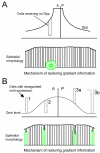
Mechanism of restoring gradient continuity. Clones and the ensuing discontinuities in Dpp signalling or Omb levels are symbolized by dotted contours. (A) Clones lacking Dpp signaling generate sharp discontinuities in the Dpp signaling gradient. Extrusion of mutant cells restores the monotonous decline of the gradient. (B) Manipulations of Omb level. Numbers next to the dotted bars refer to the experimental manipulation by which changes in Omb level were effected. (1) Upregulation of Omb in lateral tkvQD clones. (2) Loss of Omb in omb null mutant clones (strong Omb reduction by ombRNAi was similarly effective in eliciting cellular retraction). (3a) Clone of cells (tub>omb) strongly overexpressing Omb and surrounded by wild type cells. (3b) Group of wild type cells surrounded by cells strongly overexpressing Omb. In the bottom diagrams, presenting x-z views of mosaic wing discs, clonal cells are drawn in green.
Figure 7
Figure 7
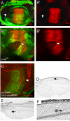
Epithelial effects of Omb misexpression are not mediated by Sal. (A, A') Strong ectopic Omb has little effect on Sal expression. The arrowhead points to where the Gal4 30 expression domain [58] overlaps the wing pouch. (B, B') Complete loss of omb (in a l(1)ombD4 clone, arrowhead) leads to strong reduction of Sal. (C) Incomplete elimination of omb function (in en>ombRNAi) only slightly decreases Sal expression. The arrowhead points to the posterior compartment, separated from the anterior compartment by a deep fold due to posterior reduction in Omb [23]. (D-F) The phenotype of omb l-o-f clones (arrows) is elicited also outside the Sal domain. Cuticular manifestations of retraction and extrusion events were found anterior to L2 (D, E) and posterior to L5 (E).

Similar articles

Cited by

References

    1. Lecuit T, Brook WJ, Ng M, Calleja M, Sun H, Cohen SM. Two distinct mechanisms for long-range patterning by Decapentaplegic in the Drosophila wing. Nature. 1996;381:387–393. doi: 10.1038/381387a0. - DOI - PubMed
    1. Nellen D, Burke R, Struhl G, Basler K. Direct and long-range action of a DPP morphogen gradient. Cell. 1996;85:357–68. doi: 10.1016/S0092-8674(00)81114-9. - DOI - PubMed
    1. Kim J, Johnson K, Chen HJ, Carroll S, Laughon A. Drosophila Mad binds to DNA and directly mediates activation of vestigial by Decapentaplegic. Nature. 1997;388:304–8. doi: 10.1038/40906. - DOI - PubMed
    1. Entchev EV, Schwabedissen A, Gonzalez-Gaitan M. Gradient formation of the TGF-beta homolog Dpp. Cell. 2000;103:981–91. doi: 10.1016/S0092-8674(00)00200-2. - DOI - PubMed
    1. Teleman AA, Cohen SM. Dpp gradient formation in the Drosophila wing imaginal disc. Cell. 2000;103:971–80. doi: 10.1016/S0092-8674(00)00199-9. - DOI - PubMed

MeSH terms