Columnar cell lesions of the canine mammary gland: pathological features and immunophenotypic analysis
- PMID: 20178635
- PMCID: PMC2841138
- DOI: 10.1186/1471-2407-10-61
Columnar cell lesions of the canine mammary gland: pathological features and immunophenotypic analysis
Abstract
Background: It has been suggested that columnar cell lesions indicate an alteration of the human mammary gland involved in the development of breast cancer. They have not previously been described in canine mammary gland. The aim of this paper is describe the morphologic spectrum of columnar cell lesions in canine mammary gland specimens and their association with other breast lesions.
Methods: A total of 126 lesions were subjected to a comprehensive morphological review based upon the human breast classification system for columnar cell lesions. The presence of preinvasive (epithelial hyperplasia and in situ carcinoma) and invasive lesions was determined and immunophenotypic analysis (estrogen receptor (ER), progesterone receptor (PgR), high molecular weight cytokeratin (34betaE-12), E-cadherin, Ki-67, HER-2 and P53) was perfomed.
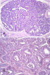
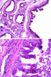
Results: Columnar cell lesions were identified in 67 (53.1%) of the 126 canine mammary glands with intraepithelial alterations. They were observed in the terminal duct lobular units and characterized at dilated acini may be lined by several layers of columnar epithelial cells with elongated nuclei. Of the columnar cell lesions identified, 41 (61.2%) were without and 26 (38.8%) with atypia. Association with ductal hyperplasia was observed in 45/67 (67.1%). Sixty (89.5%) of the columnar cell lesions coexisted with neoplastic lesions (20 in situ carcinomas, 19 invasive carcinomas and 21 benign tumors). The columnar cells were ER, PgR and E-cadherin positive but negative for cytokeratin 34betaE-12, HER-2 and P53. The proliferation rate as measured by Ki-67 appeared higher in the lesions analyzed than in normal TDLUs.
Conclusions: Columnar cell lesions in canine mammary gland are pathologically and immunophenotypically similar to those in human breast. This may suggest that dogs are a suitable model for the comparative study of noninvasive breast lesions.
Figures


Similar articles
-
Spontaneous mammary intraepithelial lesions in dogs--a model of breast cancer.Cancer Epidemiol Biomarkers Prev. 2007 Nov;16(11):2247-56. doi: 10.1158/1055-9965.EPI-06-0932. Epub 2007 Nov 2. Cancer Epidemiol Biomarkers Prev. 2007. PMID: 17982119
-
Hyperplastic and neoplastic lesions of the mammary gland in macaques.Vet Pathol. 2006 Jul;43(4):471-83. doi: 10.1354/vp.43-4-471. Vet Pathol. 2006. PMID: 16846989
-
Histological and immunohistochemical identification of atypical ductal mammary hyperplasia as a preneoplastic marker in dogs.Vet Pathol. 2012 Mar;49(2):322-9. doi: 10.1177/0300985810396105. Epub 2011 Jan 31. Vet Pathol. 2012. PMID: 21282668
-
Are columnar cell lesions the earliest histologically detectable non-obligate precursor of breast cancer?Virchows Arch. 2008 Jun;452(6):589-98. doi: 10.1007/s00428-008-0609-6. Epub 2008 Apr 24. Virchows Arch. 2008. PMID: 18437416 Review.
-
Research progress of good markers for canine mammary carcinoma.Mol Biol Rep. 2023 Dec;50(12):10617-10625. doi: 10.1007/s11033-023-08863-x. Epub 2023 Nov 9. Mol Biol Rep. 2023. PMID: 37943402 Review.
Cited by
-
Canine, Feline, and Murine Mammary Tumors as a Model for Translational Research in Breast Cancer.Vet Sci. 2025 Feb 19;12(2):189. doi: 10.3390/vetsci12020189. Vet Sci. 2025. PMID: 40005948 Free PMC article. Review.
-
Quantification of EGFR family in canine mammary ductal carcinomas in situ: implications on the histological graduation.Vet Res Commun. 2019 May;43(2):123-129. doi: 10.1007/s11259-019-09752-0. Epub 2019 Apr 24. Vet Res Commun. 2019. PMID: 31020460
-
Elevated Krüppel-like factor 4 transcription factor in canine mammary carcinoma.BMC Vet Res. 2011 Oct 7;7:58. doi: 10.1186/1746-6148-7-58. BMC Vet Res. 2011. PMID: 21978458 Free PMC article.
-
Generation of a canine anti-EGFR (ErbB-1) antibody for passive immunotherapy in dog cancer patients.Mol Cancer Ther. 2014 Jul;13(7):1777-1790. doi: 10.1158/1535-7163.MCT-13-0288. Epub 2014 Apr 22. Mol Cancer Ther. 2014. PMID: 24755200 Free PMC article.
-
Overexpression of α-enolase correlates with poor survival in canine mammary carcinoma.BMC Vet Res. 2011 Oct 21;7:62. doi: 10.1186/1746-6148-7-62. BMC Vet Res. 2011. PMID: 22014164 Free PMC article.
References
-
- Abdel-Fatah TM, Powe DG, Hodi Z, Lee AH, Reis-Filho JS, Ellis IO. High frequency of coexistence of columnar cell lesions, lobular neoplasia, and low grade ductal carcinoma in situ with invasive tubular carcinoma and invasive lobular carcinoma. Am J Surg Pathol. 2007;31(3):417–426. doi: 10.1097/01.pas.0000213368.41251.b9. - DOI - PubMed
-
- Fraser JL, Raza S, Chorny K, Connolly JL, Schnitt SJ. Columnar alteration with prominent apical snouts and secretions: a spectrum of changes frequently present in breast biopsies performed for microcalcifications. Am J Surg Pathol. 1998;22:1521–1527. doi: 10.1097/00000478-199812000-00009. - DOI - PubMed
Publication types
MeSH terms
Substances
LinkOut - more resources
Full Text Sources
Research Materials
Miscellaneous

