. 2010 Feb 19;140(4):477-90.
doi: 10.1016/j.cell.2010.01.029.

Merlin/NF2 suppresses tumorigenesis by inhibiting the E3 ubiquitin ligase CRL4(DCAF1) in the nucleus

Affiliations

Merlin/NF2 suppresses tumorigenesis by inhibiting the E3 ubiquitin ligase CRL4(DCAF1) in the nucleus

Wei Li et al. Cell. .

Abstract

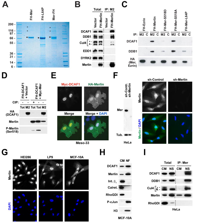
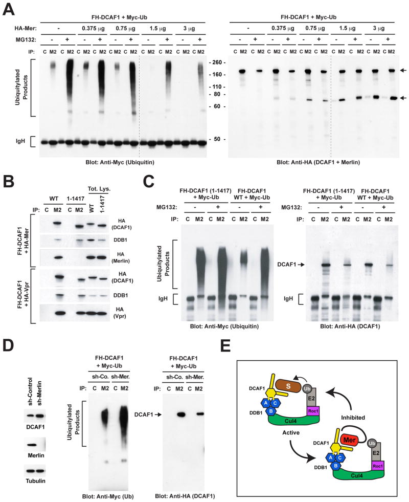
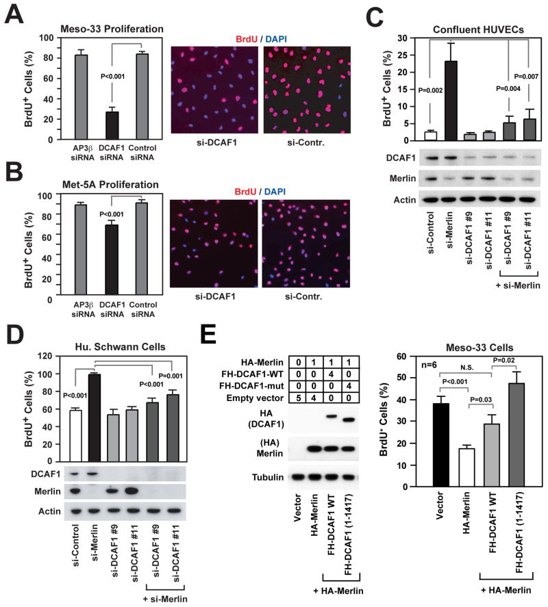
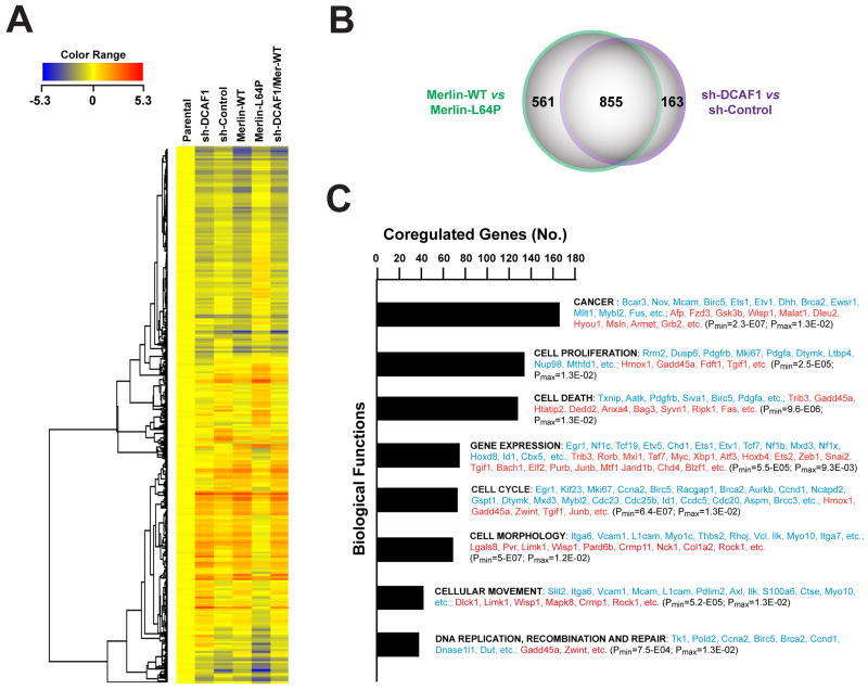
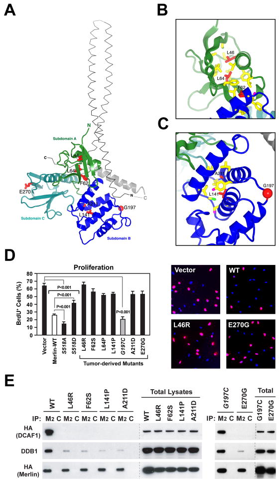
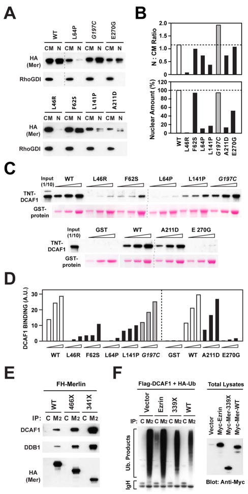
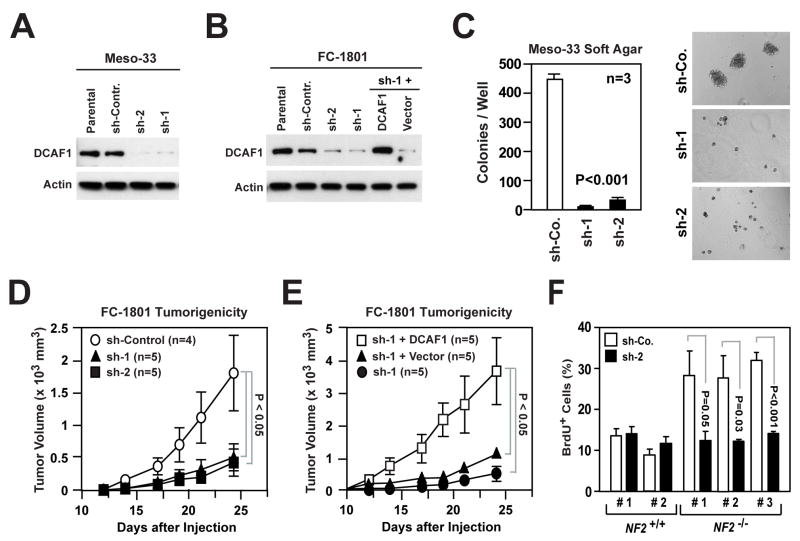
Current models imply that the FERM domain protein Merlin, encoded by the tumor suppressor NF2, inhibits mitogenic signaling at or near the plasma membrane. Here, we show that the closed, growth-inhibitory form of Merlin accumulates in the nucleus, binds to the E3 ubiquitin ligase CRL4(DCAF1), and suppresses its activity. Depletion of DCAF1 blocks the promitogenic effect of inactivation of Merlin. Conversely, enforced expression of a Merlin-insensitive mutant of DCAF1 counteracts the antimitogenic effect of Merlin. Re-expression of Merlin and silencing of DCAF1 implement a similar, tumor-suppressive program of gene expression. Tumor-derived mutations invariably disrupt Merlin's ability to interact with or inhibit CRL4(DCAF1). Finally, depletion of DCAF1 inhibits the hyperproliferation of Schwannoma cells from NF2 patients and suppresses the oncogenic potential of Merlin-deficient tumor cell lines. We propose that Merlin suppresses tumorigenesis by translocating to the nucleus to inhibit CRL4(DCAF1).


Figures

Figure 1
Figure 1. Merlin Associates with CRL4DCAF1 in the Nucleus
(A) Cos7 cells were transfected with the indicated FH-tagged (FH; FLAG-HA) forms of Merlin, lysed in RIPA, and subjected to TAP. Proteins were separated by SDS-PAGE and stained with Colloidal Blue. Dots point to the FH-tagged baits and asterisks to the 169 and 127 kD bands identified by mass spectrometry as DCAF1 and DDB1, respectively. (B) 293T cells were transfected with FH-DCAF1 or empty vector. Total lysates and Flag-immunoprecipitates (M2) were subjected to immunoblotting with antibodies to the indicated proteins. (C) As in (B), with transfection of the indicated constructs and immunoprecipitation with anti-FLAG (M2) or control (C) antibodies. (D) As in (B), with cotransfection of Myc-Merlin or empty vector and immunoprecipitation with anti-FLAG (M2) or control (C). To control for the specificity of the anti-P-Merlin antibodies, the indicated sample was treated with calf intestinal alkaline phosphatase (CIP) prior to SDS-PAGE. Endogenous Merlin was detected upon longer exposure. (E) Meso-33 cells were transiently transfected with the indicated constructs and subjected to double immunofluorescent staining with anti-HA and anti-Myc followed by DAPI. (F) HeLa cells were infected with lentiviral vectors encoding a sh-RNA targeting Merlin or a control sh-RNA and subjected to immunoblotting (left) or staining with affinity-purified antibodies to the N-terminal segment of Merlin followed by DAPI (right). (G) The indicated cells were stained as in panel F. (H) MCF-10A cells were subjected to subcellular fractionation. Equal amounts of proteins from the nuclear (NF) and non-nuclear (CM; cytosol + crude membranes) fraction were subjected to immunoblotting with antibodies to the indicated proteins. (I) Equal amounts of proteins from the non-nuclear (CM) and nuclear-soluble (NS) fraction of HeLa cells and Merlin-immunoprecipitates were immunoblotted as indicated. Anti-Cul4 recognizes Cul4-A as well as a higher molecular weight band, possibly corresponding to neddylated Cul4-A or Cul4-B (asterisk). See also Supplemental Figure S1.
Figure 2
Figure 2. Merlin Inhibits CRL4DCAF1-dependent Ubiquitylation
(A) Cos7 cells were transfected with 3 μg of FH-DCAF1 and 2 μg of Myc-Ubiquitin in combination with the indicated amounts of HA-Merlin and treated with MG132 or left untreated. Flag- or control-immunoprecipitates (M2 and C, respectively) were subjected to immunoblotting with anti-Myc to visualize ubiquitylated proteins (left) or anti-HA to visualize DCAF1 and Merlin (right). Upper arrow points to DCAF1 and lower arrow to Merlin. (B) Cos7 cells were transfected with FH-DCAF1 or FH-DCAF1 (1-1417) in combination with HA-Merlin (top panel) or HA-Vpr (bottom panel). Flag- or control-immunoprecipitates were subjected to immunoblotting. Levels of expression of DCAF1, DDB1 and Merlin or Vpr were verified by immunoblotting aliquots of total lysates. (C) As in (B), with cotransfection of Myc-Ubiquitin and treatment or not with MG132. Flag- or control-immunoprecipitates were immunoblotted with anti-Myc (left) or anti-HA (right). (D) Cos7 cells stably infected with lentiviral vectors encoding a control sh-RNA or a sh-RNA targeting Merlin were subjected to immunoblotting (left) or transfected with FH-DCAF1 and Myc-Ubiquitin followed by immunoblotting of Flag- or control-immunoprecipitates with anti-Myc (middle) or anti-HA (right). (E) Hypothetical model of Merlin-mediated inhibition of CRL4DCAF1. Merlin is proposed to function as a competitive inhibitor of CRL4DCAF1. Blue hexagons, three β propeller folds of DDB1, which contact Cul4 (B) and DCAF1 (A and C). Yellow hexagon, WD40 domain of DCAF1; yellow sticks, double DxR box implicated in binding to DDB1; S, substrate; Ub, ubiquitin; Mer, closed Merlin. See also Supplemental Figure S2.
Figure 3
Figure 3. DCAF1 Mediates Hyperproliferation and Loss of Contact Inhibition in Merlin-deficient Cells
(A, B) Meso-33 cells (A) and Met-5a cells (B) were transfected with a SMARTpool of siRNAs targeting the adaptor protein AP3β, DCAF1, or a control non-targeting pool, deprived of growth factors for 24 hours, incubated with BrdU in the presence of mitogens for 24 hours, and subjected to anti-BrdU staining followed by counterstaining with DAPI. The graph shows the percentage (± SEM) of BrdU+ cells and the pictures show representative fields. (C, D) HUVEC cells (C) and HEI 286 human Schwann cells (D) were transfected with a control si-RNA or 2 distinct si-RNAs targeting DCAF1, alone or in combination with a previously validated si-RNA targeting Merlin (Okada et al., 2005). Cells were deprived of growth factors for 24 hours, plated under confluent (C) or sparse (D) conditions on fibronectin, and subjected to BrdU incorporation in the presence of mitogens for 24 hours followed by anti-BrdU staining. The graphs show the percentage (± SEM) of BrdU+ cells. Immunoblotting was used to verify the efficiency of knock down. (E) Meso-33 cells were transiently transfected with the indicated μg of vectors encoding HA-Merlin, alone or in combination with FH-DCAF1 or FH-DCAF1 (1-1417), and subjected to immunoblotting (left) and BrdU incorporation assay (right). The anti-HA blots including FH-DCAF1 and HA-Merlin were exposed for the same time. The graph shows the percentage (±SEM) of BrdU+ cells. See also Supplemental Figure S3.
Figure 4
Figure 4. Expression of Merlin and Depletion of DCAF1 Induce a Largely Overlapping Gene Expression Program
(A) FC-1801 cells were infected with lentiviral vectors encoding a sh-RNA targeting DCAF1 (sh-DCAF1), a control sh-RNA (sh-Control), wild type Merlin, Merlin-L64P, or sh-DCAF1 in combination with wild type Merlin and subjected to DNA microarray analysis. Unsupervised clustering of 1,566 probesets differentially expressed ≥ 2 fold in at least one of the two major comparisons (sh-DCAF1 versus sh-Control and Merlin-WT versus Merlin-L64P). Values normalized to parental uninfected cells. (B) Venn Diagram shows the overlap between the list of genes differentially expressed upon expression of Merlin-WT or silencing of DCAF1 (fold change ≥ 2). Coregulated genes (885) are identified as differentially expressed ≥2 fold in one of the two comparisons and displaying a consensual significant change in the other comparison (p ≤ 0.01 by the ANOVA test). (C). 667 genes coregulated upon expression of Merlin-WT or silencing of DCAF1 (fold change ≥ 2 in both comparisons) was subjected to Ingenuity Pathway analysis. Both down-regulated (353) and upregulated genes (314) are included in the category of Biological Functions. Representative downregulated genes are shown in blue and representative upregulated genes in red in order of decreasing average fold change. See also Supplemental Figure S4.
Figure 5
Figure 5. Several Pathogenic Missense Mutations Disrupt Binding of Merlin to CRL4DCAF1 In Vivo
(A) Tumor derived mutants (red sticks) are shown in the context of the overall structure of the FERM domain of human Merlin (colored ribbons; PDB 1ISN). The α-helical coiled-coil domain from the crystal structure of the closed form of Spodoptera frugiperda Moesin (thin grey αcarbon trace; PDB 2I1K), which is highly homologous to human Merlin, is shown for reference. The coiled-coil domain was positioned by superimposing the FERM domains from the structures of Merlin and Moesin, which have a root mean square deviation for α-carbon atoms of 1.3 Å and share 62% sequence identity. (B) Close-up view of tumor-derived mutations (red sticks) located in subdomain A (green ribbon) in the crystal structure of human Merlin. Residues in contact with mutated residues are drawn as sticks and colored according to atom type (carbon, yellow; sulfur, green). (C) Subdomain B represented as in (B), with oxygens as magenta sticks. Red sphere, G197. (D) Meso-33 cells were transiently transfected with empty vector or vectors encoding wild type Merlin or indicated mutants. Asynchronized cells growing in 10% FCS were subjected to BrdU incorporation assay. The graph indicates the percentage (± SEM) of BrdU-positive cells. Pictures show representative fields of cells transfected with indicated vectors and stained with anti-BrdU (red) and DAPI (blue). (E) Cos7 cells were transfected with FH-tagged versions of wild type Merlin or indicated mutants. Flag- or control-immunoprecipitates (M2 and C, respectively) and total lysates were immunoblotted as indicated. See also Supplemental Figure S5.
Figure 6
Figure 6. Pathogenic Mutations Disrupt the Ability of Merlin to Accumulate in the Nucleus, to Bind to DCAF1, or to Inhibit CRL4DCAF1
(A) Meso-33 cells were transiently transfected with FH-tagged versions of wild type Merlin or the indicated mutants and subjected to subcellular fractionation. Equal amounts of proteins from the non-nuclear (CM) and nuclear (NF) fractions were subjected to immunoblotting. (B) The amount of Merlin or indicated mutants present in nuclear and non-nuclear fractions was estimated by densitometry of the blot in panel A. (Top) ratio of Merlin or indicated mutants present in nuclear vs. non-nuclear fractions; (Bottom) amount of each Merlin mutant present in the nucleus as a percentage over the wild type value. (C) In vitro translated and TNT-labeled DCAF1 was incubated with 0.5, 1, and 2 μg of GST fusion proteins comprising the FERM domain of wild type Merlin or indicated mutants. GST-bound DCAF1 was detected by blotting with streptavidin. (D) Densitometric analysis of (C). The graph shows the binding values in arbitrary units. (E) Cos7 cells were transfected with the indicated FH-tagged forms of Merlin and subjected to immunoprecipitation with control (C) or anti-Flag Mab (M2) followed by immunoblotting. (F) Cos7 cells were transfected with FH-DCAF1 and HA-Ubiquitin in combination with the indicated Myc-tagged constructs or empty vector and treated with MG132. Flag- or control-immunoprecipitates were immunoblotted with anti-HA (left). Total lysates were immunoblotted with anti-Myc (right).
Figure 7
Figure 7. Silencing of DCAF1 Suppresses the Tumorigenic Potential of Merlin-deficient Cells
(A) Meso-33 cells were infected with lentiviral vectors encoding two distinct sh-RNAs targeting DCAF1 (sh-1 and sh-2) or a control sh-RNA (sh-Contr.) and subjected to immunoblotting. (B) FC-1801 cells treated as in (A). Cells infected with sh-1 were also infected with a retroviral vector encoding a sh-RNA-resistant version of DCAF1 or with empty vector. Immunoblotting was as indicated. (C) Meso-33 cells infected with the indicated lentiviral vectors were subjected to soft agar assay. The graph illustrates the total number (± SEM) of colonies present in each well after 2 weeks of culture. Representative images shown. (D, E) 2 × 10⁶ FC-1801 cells infected with the indicated lentiviral vectors were injected subcutaneously in nude mice. (D) shows the increase in volume (± SEM) over time of tumors generated by cells infected with lentiviruses encoding a control sh-RNA (sh-Control) or two distinct sh-RNAs targeting DCAF1 (sh-1 and sh-2). (E) shows the increase in volume (± SEM) over time of tumors generated by cells infected with sh-1 and either a retroviral vector encoding a sh-RNA-resistant version of DCAF1 or empty vector. Data were obtained from the same experiment. Note the different scale of the Y axes of the two graphs. (F) Primary Schwann cells from 2 normal individuals (NF2+/+) and Schwannoma cells from 2 NF2 patients (NF2−/−) were infected with lentiviruses encoding a sh-RNA targeting DCAF1 (sh-2) or a control sh-RNA (sh-Co.), deprived of growth factors, restimulated with mitogens in the presence of BrdU, and subjected to anti-BrdU staining. The graph shows the percentage (± SEM) of BrdU+ cells. See also Supplemental Figure S6.

Similar articles

Cited by

References

    1. Ahronowitz I, Xin W, Kiely R, Sims K, MacCollin M, Nunes FP. Mutational spectrum of the NF2 gene: a meta-analysis of 12 years of research and diagnostic laboratory findings. Hum Mutat. 2007;28:1–12. - PubMed
    1. Ammoun S, Flaiz C, Ristic N, Schuldt J, Hanemann CO. Dissecting and targeting the growth factor-dependent and growth factor-independent extracellular signal-regulated kinase pathway in human schwannoma. Cancer Res. 2008;68:5236–5245. - PubMed
    1. Bretscher A, Edwards K, Fehon RG. ERM proteins and merlin: integrators at the cell cortex. Nat Rev Mol Cell Biol. 2002;3:586–599. - PubMed
    1. Cho E, Feng Y, Rauskolb C, Maitra S, Fehon R, Irvine KD. Delineation of a Fat tumor suppressor pathway. Nat Genet. 2006;38:1142–1150. - PubMed
    1. Clevers H. Wnt/beta-catenin signaling in development and disease. Cell. 2006;127:469–480. - PubMed

Publication types

Associated data