. 2010 Feb 19;140(4):504-16.
doi: 10.1016/j.cell.2010.01.019.

Dicer-independent primal RNAs trigger RNAi and heterochromatin formation

Affiliations

Dicer-independent primal RNAs trigger RNAi and heterochromatin formation

Mario Halic et al. Cell. .

Abstract

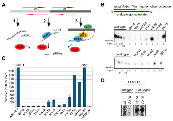
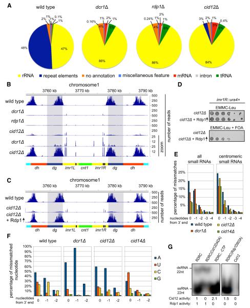
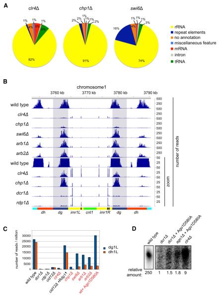
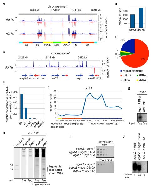
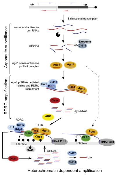
Assembly of fission yeast pericentromeric heterochromatin and generation of small interfering RNAs (siRNAs) from noncoding centromeric transcripts are mutually dependent processes. How this interdependent positive feedback loop is first triggered is a fundamental unanswered question. Here, we show that two distinct Argonaute (Ago1)-dependent pathways mediate small RNA generation. RNA-dependent RNA polymerase complex (RDRC) and Dicer act on specific noncoding RNAs to generate siRNAs by a mechanism that requires the slicer activity of Ago1 but is independent of pre-existing heterochromatin. In the absence of RDRC or Dicer, a distinct class of small RNAs, called primal small RNAs (priRNAs), associates with Ago1. priRNAs are degradation products of abundant transcripts, which bind to Ago1 and target antisense transcripts that result from bidirectional transcription of DNA repeats. Our results suggest that a transcriptome surveillance mechanism based on random association of RNA degradation products with Argonaute triggers siRNA amplification and heterochromatin assembly within DNA repeats.


Figures

Figure 1
Figure 1. Detection of Small RNAs in RNAi and Heterochromatin Mutant Backgrounds with Splinted Ligation
(A) Possible models for biogenesis of initial siRNAs from centromeric repeats, including dsRNA resulting from bidirectional transcription (1), RDRC recognition of specific sequences (2), and RITS/RDRC recruitment by pre-existing H3K9 methylation (3). (B) Detection of Argonaute-associated centromeric small RNAs in wild-type and the indicated mutant cells by splinted ligation. Bridge oligonucleotides corresponding to eight different centromeric siRNAs mapping to both dg and dh transcripts were used. Note that different amounts of Ago1-associated small RNAs, indicated as relative input, were used for different strains and normalized to the levels in wild-type cells. Note different exposure times for the upper and lower panels. (C) Quantification of the splinted ligation data by Quantity One software. Splinted ligation experiments were performed with three or more biological replicates, and standard deviations are shown. Small RNA quantification for dis3-54 was done twice, and error bars represent deviation from the mean. rrp6Δ and cid14Δ data are based on only one experiment and are consistent with previous findings. The signal for dcr1Δ was set to 1 and enrichment over dcr1Δ is shown. (D) Detection of Argonaute-associated centromeric small RNAs by splinted ligation in dcr1Δ and rdp1Δ cells. In untagged purifications from wild-type and dcr1Δ cells no centromeric small RNAs were detected. See also Figure S1.
Figure 2
Figure 2. Roles of Dicer and RDRC in Small RNA Generation
(A) S. pombe Ago1-associated small RNAs were analyzed by high-throughput sequencing from wild-type, dcr1Δ, rdp1Δ, and cid12Δ cells, and classified as indicated below the pie charts. Pie charts illustrate percentages for the individual small RNA classes relative to the total number of small RNA reads for each strain. (B) Small RNA reads in wild-type and the indicated mutant cells were plotted over centromeric repeat region of chromosome 1. The location of the centromere (cnt1, green), the imr repeats (imr1L and imr1R, yellow), tRNAs (black), the dg (blue), and dh (red) repeats are indicated below the siRNA peaks. Chromosome coordinates are indicated above the peaks. Scale bars on the right denote small RNA reads numbers normalized per one million reads. (C) Small RNA reads in wild-type, cid12Δ and cid12Δ overexpressing Rdp1. Shaded areas highlight the dg repeats. (D) Silencing assay showing that Rdp1 overexpression rescues the cid12Δ loss of silencing phenotype of a imr1R::ura4+ reporter gene. Plating efficiency and silencing are assayed by growth on complete selective (EMMC-Leu) and 5-Fluoroorotic acid (FOA) medium, respectively. (E) Percentage of mismatched nucleotides for all small RNAs (sRNAs) and centromeric small RNAs in wild-type, dcr1Δ, cid12Δ and cid14Δ cells. The last 5 nt are shown from 3′ end of small RNA. 0 denotes the last nucleotide, —1 the nucleotide prior to the last. (F) Identity of mismatched nucleotides in small RNAs in wild-type, dcr1Δ, cid12Δ, and cid14Δ cells. As in (E), mismatched nucleotides are shown from 3′ end of small RNAs. (G) Autoradiograph of native polyacrylamide gel showing the Cid12 and Rdp1 activities on a 22 nt single-stranded RNA (ssRNA) template using 32P-ATP incorporation. Cid12 products migrate as ssRNA, whereas Rdp1 products migrate as double-stranded RNA (dsRNA). No Rdp1 dsRNA synthesis activity was detectable in absence of CTP or with the catalytically inactive Rdp1-D903A enzyme. See also Figure S2.
Figure 3
Figure 3. Heterochromatin-Independent siRNA Generation from dg Transcripts
(A) High-throughput sequencing of small RNAs from the clr4Δ, chp1Δ, and swi6Δ cells. Pie charts illustrate percentages for the individual small RNA classes relative to the total number of small RNA reads for each strain, classified as indicated. (B) Small RNA reads from wild-type and the indicated mutant cells are plotted over centromeric repeat region of chromosome 1, at 10× zoom. (The lower part of panel B shows that in chp1Δ and clr4Δ cells, dg siRNAs levels are much higher than dh siRNA levels.) (C) Number of small RNA reads mapping to dg1L and dh1L in indicated cells. Small RNA read numbers were normalized per one million reads. (D) Splinted ligation detection of small RNAs associated with Ago1 and Ago1D580A in wild-type, dcr1Δ, ago1Δ, and clr4Δ cells. Five different centromeric small RNAs mapping to dg transcripts were assayed. See also Figure S3.
Figure 4
Figure 4. priRNAs Primarily Target the Centromeric Repeats and Require the PAZ/MID Domains for Binding to Ago1
(A) Small RNA reads from dcr1Δ and rdp1Δ cells are plotted over centromeric region on chromosome 1 in a strand specific way (blue, + strand; red, −strand). Small RNAs in dcr1Δ cells are referred to as priRNAs. (B) Number of centromeric small RNA reads in dcr1Δ and rdp1Δ cells. Small RNA read numbers were normalized per one million reads. (C) Small RNA densities from dcr1Δ are plotted over a selected euchromatic region on chromosome 1 in a strand-specific way. Note that higher number of reads is often present after the 3′ end of mRNA transcripts. (D) Classification of antisense priRNAs, as indicated next to the pie charts. Pie charts illustrate the percentages of the individual priRNA classes relative to the total number of antisense priRNAs. Twenty-two percent of antisense priRNAs map to repeat elements. (E) Antisense small RNA reads for each class are normalized to the number of transcripts they may target. Antisense priRNAs target mainly centromeric dg and dh transcripts. (F) Average genome-wide distribution of priRNAs mapping to chromosome 1 mRNAs in dcr1Δ cells. The coding region of each mRNA was divided to five equal parts, each representing 20% of the protein coding sequence. The 3′ untranslated region (UTR) and downstream regions were divided into windows of 50 and 100 nucleotides. The number of priRNA reads mapping to each window was normalized to the size of the corresponding window and to the total number of mRNAs. The number of priRNA reads represents normalized average number of reads per 1 kb window. (G) Splinted ligation detection of centromeric small RNAs associated with Argonaute or from total RNA, showing that priRNAs are enriched in the Argonaute purification. (H) Sybr Green II staining of 17.5% denaturing polyacrylamide gel with 3 μg of RNAs associated with FLAG-Ago1 or FLAG-Ago1-3A purified from dcr1Δ cells. RNAs of approximately 22 nt in length are specifically associated with wild-type FLAG-Ago1. (I) Silencing assay showing that the expression of wild-type Ago1, but not Ago1-D580A or Ago1-3A (F276A/Y513A/K517A) mutants, rescues loss of silencing in ago1Δ cells. Growth on FOA medium indicates ura4+ silencing. (J) Splinted ligation detection of small RNAs associated with wild-type and mutant Ago1 proteins, showing that Ago1-3A containing three point mutations in the MID and PAZ domains abolishes small RNA binding. Ago1-F276A containing a single point mutation in the PAZ domain binds small RNAs more weakly. See also Figure S4.
Figure 5
Figure 5. Role for priRNAs in Promoting Histone H3K9 Methylation
(A) Location of PCR products in ChIP and qRT-PCR experiments are indicated as white bars below the dg (blue), dh (red), and imr1L (yellow) regions of chromosome 1. Green bars depict regions with mapped siRNAs. (B) Chromatin immunoprecipitation (ChIP) experiments showing Dcr1-independent and Ago1-dependent H3K9 methylation at the indicated centromeric dgG region (also see Figure S5). ChIP experiments were performed with H3K9me2 antibody. Relative fold enrichment over clr4Δ is indicated bellow each panel. (C) ChIP experiments showing that priRNA/siRNAs can induce low level of H3K9 methylation independently of the Chp1 and Tas3 subunits of RITS. Ago1-3A mutant deficient in priRNA binding is unable to direct H3K9 methylation. (D) ChIP experiments showing that slicer activity of Ago1 is essential for priRNA-dependent H3K9 methylation; serial dilutions containing 1 and 1/30th μl of immunoprecipitated DNA were used and the lower dilution, for which PCR is in the linear range for all samples, was used for quantification. (E) Quantification of the ChIP data. ChIP experiments were performed several times with independent biological replicates and average values with standard deviation are shown as enrichment over clr4Δ. The average enrichment for wild-type, dcr1Δ, and ago1Δ was based on more than 15 experiments with different primers sets. p values were calculated using the Student’s t test. (F and G) Increase in priRNA levels correlates with increased H3K9 methylation. Splinted ligation detection of Argonaute-associated centromeric small RNAs showing that centromeric priRNAs are 3.5-fold more abundant in dcr1Δcid14Δ than in dcr1Δ cells (F). Splinted ligation was performed as described in Figure 1. Quantification of ChIP experiments showing 2-fold higher levels of H3K9 methylation in dcr1Δcid14Δ than in dcr1Δ. The dgB primers were used in this ChIP experiments and the results show the average for seven independent biological replicates. Error bars indicate standard deviations. p value was calculated using the Student’s t test. See also Figure S5.
Figure 6
Figure 6. Ago1-Bound siRNAs Are Trimmed after Release of the Passenger Strand
(A) Small RNA reads in wild-type cells, with and without expression of the Ago1D580A dominant negative mutant, are plotted over centromeric repeat region of chromosome 1. (B) Length distribution of centromeric siRNAs associated with Ago1 and Ago1-D580A, showing the broader siRNA length distribution of Ago1-D580A-associated siRNAs. (C) Centromeric siRNAs with identical 5′ ends and different 3′ ends bound by Ago1 and Ago1-D580A. The number of reads and the length are indicated for each siRNA. Mismatched residues are shown in underlined blue. (D) Length distribution of centromeric siRNAs from (C) with identical 5′ ends bound by Ago1 and Ago1-D580A. The siRNA is extended at its 3′ end in Ago1-D580A, suggesting that in wild-type Ago1 the siRNA is trimmed from its 3′ end. (E) Length distribution of centromeric dg siRNAs that perfectly match the S. pombe genome associated with Ago1-D580A compared to the wild-type. (F) Splinted ligation detection of Argonaute-associated centromeric small RNAs in indicated mutant cells. The levels of centromeric priRNAs are similar in dcr1Δrdp1Δ, dcr1Δeri1Δ and dcr1ΔDis3-54,D171N mutant cells. Splinted ligation detection was performed as described in Figure 1. (G) Number of priRNA reads normalized to one million reads mapping to dg in dcr1Δ and dcr1Δdis3-54,D171N mutant cells. priRNAs are approximately 2-fold more abundant in dcr1Δ than in dcr1Δdis3-54,D171N mutant cells. (H) Size distribution of centromeric siRNAs associated with Ago1 in wild-type and dis3-54,D171N mutant cells. Approximately 3% of total siRNAs have a larger average size in dis3-54,D171N mutant cells compared to the wild-type cells. See also Figure S6.
Figure 7
Figure 7. Model for Initiation of siRNA Generation and Heterochromatin Assembly
Model summarizing pri/siRNA generation and amplification in heterochromatin-independent and heterochromatin-dependent manners. priRNAs are generated by degradation of centromeric transcripts and loaded onto Ago1. priRNAs target centromeric transcripts and induce heterochromatin independent siRNA generation in an Ago1 slicer- and RDRC/Dicer-dependent manner. Amplified siRNAs target nascent transcripts, recruit RITS, RDRC, and CLRC complexes to induce efficient siRNA amplification and H3K9 methylation. The ARC complex is required for this heterochromatin-dependent amplification step. priRNAs can also target chromatin bound nascent centromeric transcripts (dashed arrow), recruit the CLRC complex, and induce low levels of H3K9 methylation generation. The Cid12 and Cid14 nucleotidyltransferase enzymes modify the 3′ ends of small RNAs, leading to exosome-mediated trimming or degradation. CLRC, Clr4 H3K9 methyltransferase complex; black lollipops, H3K9 methylation.

Comment in

References

    1. Aoki K, Moriguchi H, Yoshioka T, Okawa K, Tabara H. In vitro analyses of the production and activity of secondary small interfering RNAs in C. elegans. EMBO J. 2007;26:5007–5019. - PMC - PubMed
    1. Aravin AA, Hannon GJ, Brennecke J. The Piwi-piRNA pathway provides an adaptive defense in the transposon arms race. Science. 2007;318:761–764. - PubMed
    1. Baulcombe D. RNA silencing in plants. Nature. 2004;431:356–363. - PubMed
    1. Bühler M, Verdel A, Moazed D. Tethering RITS to a nascent transcript initiates RNAi- and heterochromatin-dependent gene silencing. Cell. 2006;125:873–886. - PubMed
    1. Bühler M, Haas W, Gygi SP, Moazed D. RNAi-dependent and -independent RNA turnover mechanisms contribute to heterochromatic gene silencing. Cell. 2007;129:707–721. - PubMed

Publication types

MeSH terms

Associated data