

A differential cell capture assay for evaluating antibody interactions with cell surface targets
- PMID: 20178770
- PMCID: PMC4485450
- DOI: 10.1016/j.ab.2010.02.015
A differential cell capture assay for evaluating antibody interactions with cell surface targets
Abstract
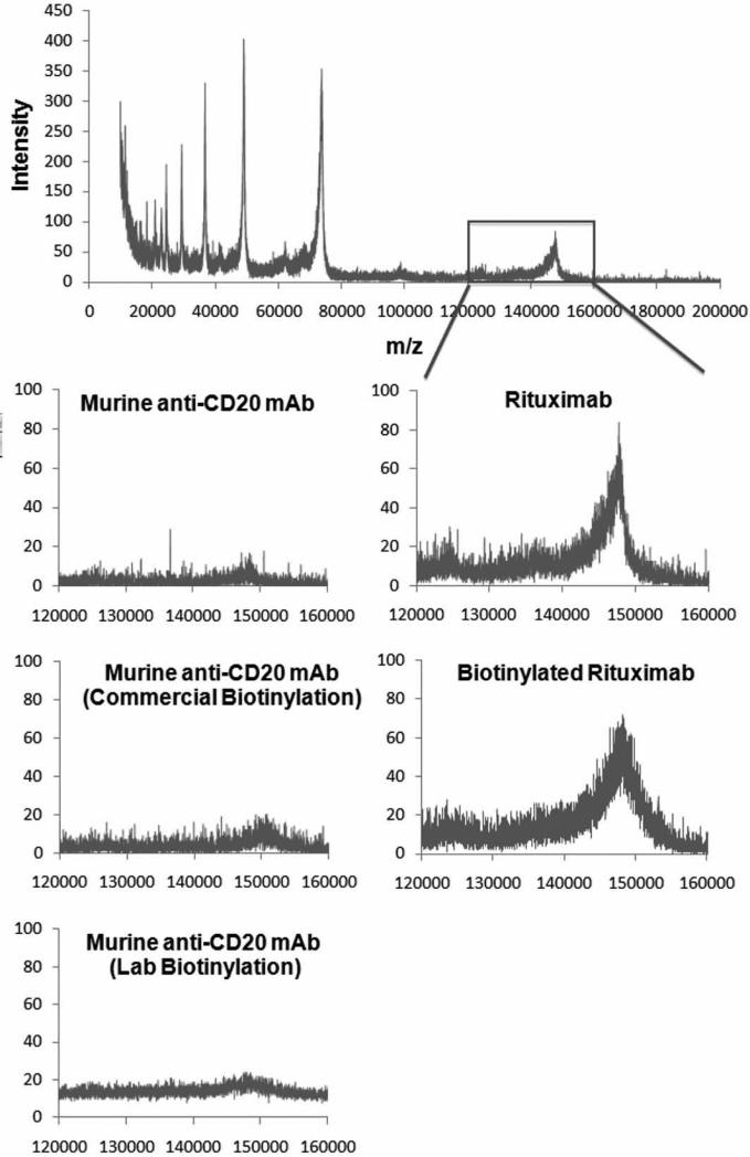
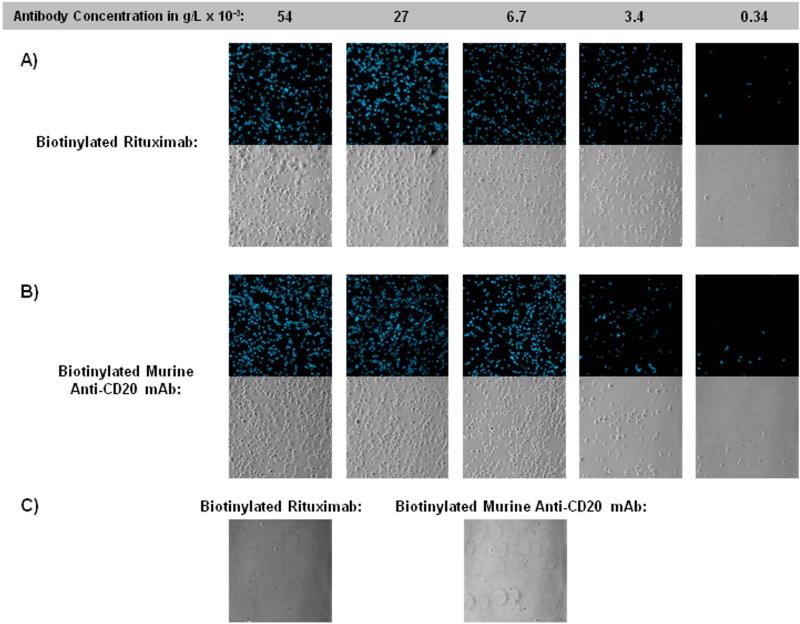
Many biological and biomedical laboratory assays require the use of antibodies and antibody fragments that strongly bind to their cell surface targets. Conventional binding assays, such as the enzyme-linked immunosorbent assay (ELISA) and flow cytometry, have many challenges, including capital equipment requirements, labor intensiveness, and large reagent and sample consumption. Although these techniques are successful in mainstream biology, there is an unmet need for a tool to quickly ascertain the relative binding capabilities of antibodies/antibody fragments to cell surface targets on the benchtop at low cost. We describe a novel cell capture assay that enables several candidate antibodies to be evaluated quickly as to their relative binding efficacies to their cell surface targets. We used chimeric rituximab and murine anti-CD20 monoclonal antibodies as cell capture agents on a functionalized microscope slide surface to assess their relative binding affinities based on how well they capture CD20-expressing mammalian cells. We found that these antibodies' concentration-dependent cell capture profiles correlate with their relative binding affinities. A key observation of this assay involved understanding how differences in capture surfaces affect the assay results. This approach can find utility when an antibody or antibody fragment against a known cell line needs to be selected for targeting studies.
Published by Elsevier Inc.
Figures


Similar articles
-
Expression and biological characterization of an anti-CD20 biosimilar candidate antibody: a case study.MAbs. 2012 Jul-Aug;4(4):488-96. doi: 10.4161/mabs.20761. Epub 2012 Jul 1. MAbs. 2012. PMID: 22647435 Free PMC article.
-
Binding to CD20 by anti-B1 antibody or F(ab')(2) is sufficient for induction of apoptosis in B-cell lines.Cancer Immunol Immunother. 2002 Mar;51(1):15-24. doi: 10.1007/s00262-001-0247-1. Epub 2001 Dec 18. Cancer Immunol Immunother. 2002. PMID: 11845256 Free PMC article.
-
γδ T-cell killing of primary follicular lymphoma cells is dramatically potentiated by GA101, a type II glycoengineered anti-CD20 monoclonal antibody.Haematologica. 2011 Mar;96(3):400-7. doi: 10.3324/haematol.2010.029520. Epub 2010 Nov 25. Haematologica. 2011. PMID: 21109686 Free PMC article.
-
CD20-targeted therapy: the next generation of antibodies.Semin Hematol. 2010 Apr;47(2):199-210. doi: 10.1053/j.seminhematol.2010.01.007. Semin Hematol. 2010. PMID: 20350667 Review.
-
The clinical and biological role of CD20 membrane antigen modulation under immunotherapy with anti-CD20 monoclonal antibody rituximab in lymphoprolipherative neoplastic disorders.Expert Opin Biol Ther. 2011 May;11(5):551-7. doi: 10.1517/14712598.2011.567262. Epub 2011 Mar 9. Expert Opin Biol Ther. 2011. PMID: 21385115 Review.
Cited by
-
A microfluidic cell concentrator.Anal Chem. 2010 Oct 1;82(19):8320-6. doi: 10.1021/ac101866p. Anal Chem. 2010. PMID: 20843010 Free PMC article.
-
Characterization of cell seeding and specific capture of B cells in microbubble well arrays.Biomed Microdevices. 2013 Jun;15(3):453-63. doi: 10.1007/s10544-013-9745-0. Biomed Microdevices. 2013. PMID: 23358874 Free PMC article.
-
Point-of-care (POC) devices by means of advanced MEMS.Talanta. 2015 Dec 1;145:55-9. doi: 10.1016/j.talanta.2015.04.032. Epub 2015 Apr 23. Talanta. 2015. PMID: 26459443 Free PMC article. Review.
References
-
- Wu AM, Senter PD. Arming antibodies: prospects and challenges for immunoconjugates. Nat. Biotechnol. 2005;23:1137–1146. - PubMed
-
- Filpula D. Antibody engineering and modification technologies. Biomol. Eng. 2007;24:201–215. - PubMed
-
- Baselga J, Norton L, Albanell J, Kim Y-M, Mendelsohn J. Recombinant humanized anti-HER2 antibody (HerceptinTM) enhances the antitumor activity of Paclitaxel and Doxorubicin against HER2/neu overexpressing human breast cancer xenografts. Cancer Res. 1998;58:2825–2831. - PubMed
-
- Wong JYC, Chu DZ, Williams LE, Yamauchi DM, Ikle DN, Kwok CS, Liu A, Wilczynski S, Colcher D, Yazaki PJ, Shively JE, Wu AM, Raubitschek AA. Pilot trial evaluating an 123I-labeled 80-kilodalton engineered anticarcinoembryonic antigen antibody fragment (cT84.66 Minibody) in patients with colorectal cancer. Clin. Cancer Res. 2004;10:5014–5021. - PubMed
-
- Jiao J, Wang S, Qiao R, Vivanco I, Watson PA, Sawyers CL, Wu H. Murine cell lines derived from Pten null prostate cancer show the critical role of PTEN in hormone refractory prostate cancer development. Cancer Res. 2007;67:6083–6091. - PubMed
Publication types
MeSH terms
Substances
Grants and funding
LinkOut - more resources
Full Text Sources
Other Literature Sources
