Review
. 2010 May 17;584(10):2033-42.
doi: 10.1016/j.febslet.2010.02.045. Epub 2010 Feb 20.

Large conductance, Ca2+-activated K+ channels (BKCa) and arteriolar myogenic signaling

Affiliations
Review

Large conductance, Ca2+-activated K+ channels (BKCa) and arteriolar myogenic signaling

Michael A Hill et al. FEBS Lett. .

Abstract

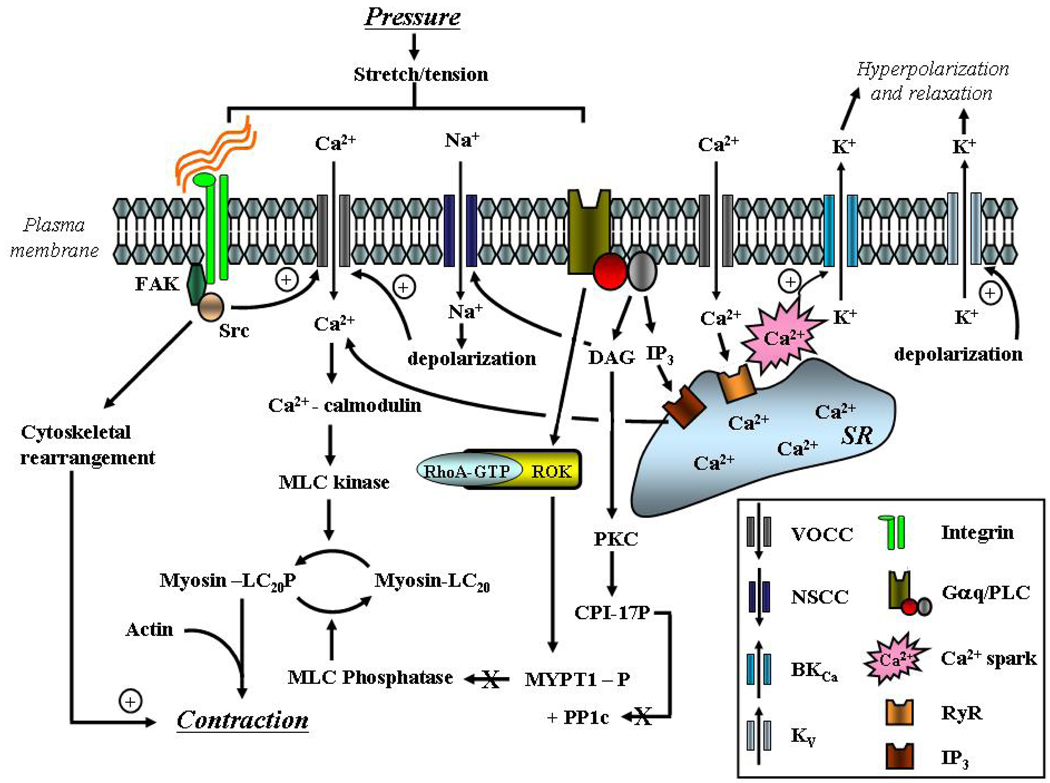
Myogenic, or pressure-induced, vasoconstriction is critical for local blood flow autoregulation. Underlying this vascular smooth muscle (VSM) response are events including membrane depolarization, Ca(2+) entry and mobilization, and activation of contractile proteins. Large conductance, Ca(2+)-activated K(+) channel (BK(Ca)) has been implicated in several of these steps including, (1) channel closure causing membrane depolarization, and (2) channel opening causing hyperpolarization to oppose excessive pressure-induced vasoconstriction. As multiple mechanisms regulate BK(Ca) activity (subunit composition, membrane potential (Em) and Ca(2+) levels, post-translational modification) tissue level diversity is predicted. Importantly, heterogeneity in BK(Ca) channel activity may contribute to tissue-specific differences in regulation of myogenic vasoconstriction, allowing local hemodynamics to be matched to metabolic requirements. Knowledge of such variability will be important to exploiting the BK(Ca) channel as a therapeutic target and understanding systemic effects of its pharmacological manipulation.


Figures

Figure 1
Figure 1
Schematic diagram illustrating the subunit structure of BKCa (upper panel) and the activation of BKCa through SR-mediated Ca2+ sparks (lower panel). Upper Panel: in VSM cells BKCa exists as a tetramer of α subunits arranged around a central conducting pore. The α subunit has 7 membrane spanning domains and an intracellular C-terminal tail containing a number of regulatory sites (see text for detail). Co-assembled with each α subunit is a β1 subunit, which confers additional Ca2+ and voltage sensitivity on the channel. Lower Panel: close apposition of the SR and plasma membranes creates a restricted space in which Ca2+ can increase to levels required for activation of BKCa. Ca2+ increases occur transiently in the form of Ca2+ sparks due to periodic release from the SR via ryanodine-sensitive mechanisms.
Figure 2
Figure 2
Schematic diagram illustrating signaling mechanisms underlying arteriolar myogenic vasoconstriction. Emphasis is placed on ion channels, membrane depolarization, mobilization of Ca2+ and activation of the contractile proteins. In addition, negative feedback activation of BKCa by SR-mediated Ca2+ sparks is shown (from Hill et al.[124]).
Figure 3
Figure 3
Characteristics of STOCs in VSM cells enzymatically isolated from cremaster muscle arterioles and small cerebral arteries. The upper panels show example STOC tracings and illustrate that despite differences in apparent voltage dependency, ryanodine eliminates the outward currents in both cell preparations. Recordings were made at a pipette [Ca2+] of 100 nM. The lower panel shows that at any given holding potential STOCs are both less frequent and of smaller amplitude in cremaster VSM cells compared to those from cerebral arteries. Data are re-plotted from Yan et al and are shown as mean SEM for n = 6 – 8 cells.

Similar articles

Cited by

References

    1. Jackson WF. Potassium channels in the peripheral microcirculation. Microcirculation. 2005;12:113–127. - PMC - PubMed
    1. Feletou M. Calcium-activated potassium channels and endothelial dysfunction: therapeutic options? Br J Pharmacol. 2009;156:545–562. - PMC - PubMed
    1. Feletou M, Vanhoutte PM. EDHF: an update. Clin Sci (Lond) 2009;117:139–155. - PubMed
    1. Liu Y, Pleyte K, Knaus HG, Rusch NJ. Increased expression of Ca2+-sensitive K+ channels in aorta of hypertensive rats. Hypertension. 1997;30:1403–1409. - PubMed
    1. McGahon MK, et al. Diabetes downregulates large-conductance Ca2+-activated potassium beta 1 channel subunit in retinal arteriolar smooth muscle. Circ Res. 2007;100:703–711. - PMC - PubMed

Publication types

MeSH terms

Substances