. 2010 May 7;285(19):14791-805.
doi: 10.1074/jbc.M110.116905. Epub 2010 Feb 23.

cAMP/CREB-mediated transcriptional regulation of ectonucleoside triphosphate diphosphohydrolase 1 (CD39) expression

Affiliations

cAMP/CREB-mediated transcriptional regulation of ectonucleoside triphosphate diphosphohydrolase 1 (CD39) expression

Hui Liao et al. J Biol Chem. .

Abstract

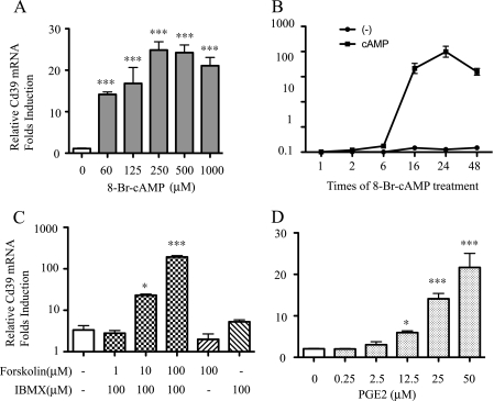
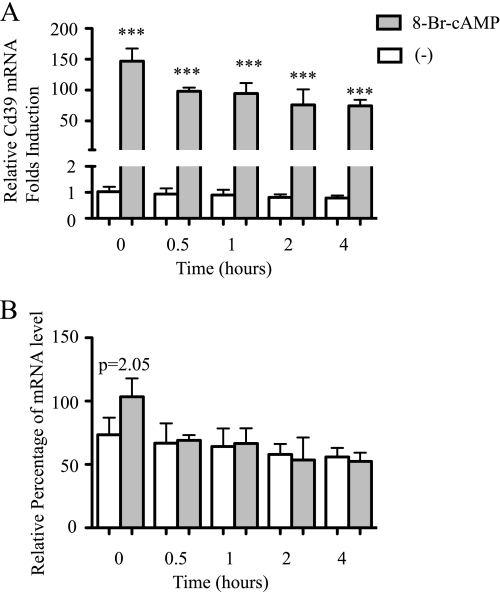
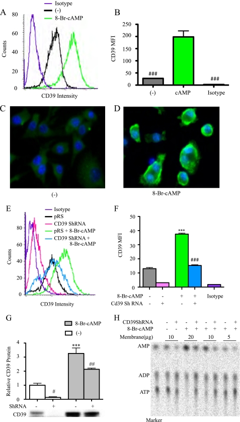
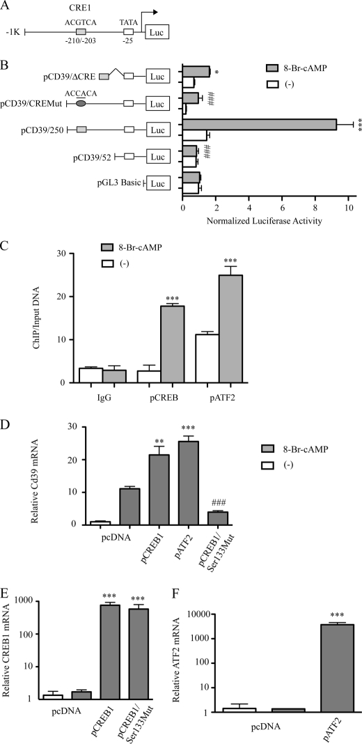
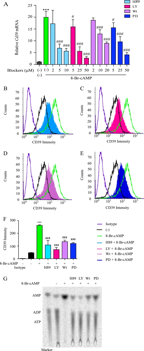
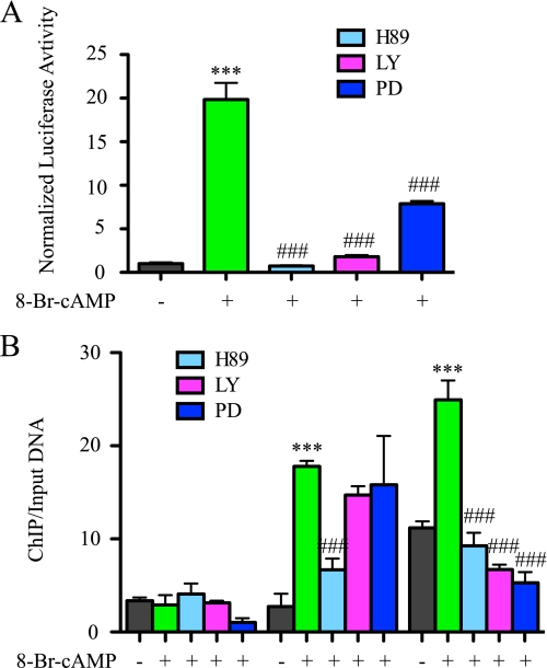
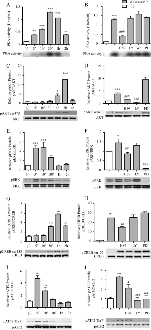
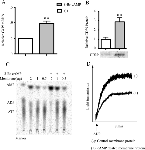
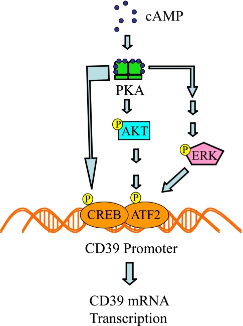
CD39 is a transmembrane enzyme that inhibits platelet reactivity and inflammation by phosphohydrolyzing ATP and ADP to AMP. Cyclic AMP (cAMP), an essential second messenger, is particularly important in regulating genes controlling vascular homeostasis. These experiments test the hypothesis that cAMP might positively regulate the expression of CD39 and thereby modulate important vascular homeostatic properties. Cd39 mRNA was induced by 13.8- fold in RAW cells treated with a membrane-permeant cAMP analogue (8-bromo-cyclic AMP; 8-Br-cAMP), stimulation of adenylate cyclase, or prostanoids known to drive cAMP response. Fluorescence-activated cell sorting, immunofluorescence, and TLC assays demonstrated that both CD39 protein expression and enzymatic activity were increased in cells treated with 8-Br-cAMP but not in cells transfected with short hairpin RNA against CD39. This analogue drove a significant increase in transcriptional activity at the Cd39 promoter although not when the promoter's cAMP-response element sites were mutated. Pretreatment with cAMP-dependent protein kinase (PKA), phosphoinositide 3-kinase (PI3K), or ERK inhibitors nearly obliterated the cAMP-driven increase in Cd39 mRNA, protein expression, and promoter activity. 8-Br-cAMP greatly increased the phosphorylation of CREB1 (Ser(133)) and ATF2 (Thr(71)) in a PKA-, PI3K-, and ERK-dependent fashion. Chromatin immunoprecipitation assays demonstrated that binding of phosphorylated CREB1 and ATF2 to cAMP-response element-like sites was significantly increased with 8-Br-cAMP treatment and that binding was reduced with PKA, PI3K, and ERK inhibition, whereas transfection of Creb1 and Atf2 overexpression constructs enhanced cAMP-driven Cd39 mRNA expression. Transfection of RAW cells with mutated Creb1 (S133A) reduced cAMP-driven Cd39 mRNA expression. Furthermore, the cAMP-mediated induction of Cd39 mRNA, protein, and phosphohydrolytic activity was replicated in primary peritoneal macrophages. These data identify cAMP as a crucial regulator of macrophage CD39 expression and demonstrate that cAMP acts through the PKA/CREB, PKA/PI3K/ATF2, and PKA/ERK/ATF2 pathways to control a key vascular homeostatic mediator.


Figures

FIGURE 1.
FIGURE 1.
Effect of cAMP on macrophage Cd39 mRNA expression. RAW cells were treated with 8-Br-cAMP for the indicated times and dosages, and total RNA was isolated. qRT-PCR was used to detect Cd39 mRNA expression in dose- and time-dependent manners (A and B). RAW cells were treated with forskolin and isobutylmethylxanthine or PGE2 at the indicated dosages for 16 h, and total RNA was isolated. Quantitative RT-PCR was used to detect Cd39 mRNA expression (C and D). *, p < 0.05; ***, p < 0.001 compared with non-treatment controls. ###, p < 0.001 compared with maximum Cd39 mRNA expression.
FIGURE 2.
FIGURE 2.
Effect of cAMP on Cd39 mRNA stability in macrophages. RAW cells were treated with 8-Br-cAMP for 4 h and then incubated with 5 μg/ml of actinomycin D for the indicated times. Total RNA was isolated, and then qRT-PCR was used to detect Cd39 mRNA expression as described under “Materials and Methods.” The values of RQ were normalized to the values of RQ for the 0 min actinomycin D-treated control (non-cAMP-treated) cells (A). In order to compare the relative rates of RNA degradation, the values of RQ for actinomycin D and cAMP co-treated cells were normalized to the 0 min actinomycin D and cAMP-treated cells and shown as percentages (B). In order to assess the relative levels of Cd39 mRNA, the values of actinomycin D and non-cAMP treated samples were normalized to C3 (0 min actinomycin D-treated normoxic cells) (B) (26). ***, p < 0.001. 0, 0.5, 1, 2, or 4 h of 8-Br-cAMP treatment versus non-cAMP-treated cells, respectively.
FIGURE 3.
FIGURE 3.
Effect of cAMP on macrophage CD39 protein expression. RAW cells were treated with 250 μm 8-Br-cAMP for 16 h, and CD39 protein expression was measured by FACS (Isotype, rat IgG2a isotype control; MFI, mean fluorescence intensity) (A and B) and immunofluorescence staining (C and D). RAW cells were stably transfected with Cd39 shRNA or pRS shRNA control vector. The transfected cells were treated with 250 μm 8-Br-cAMP for 5 h, CD39 protein expression was measured by FACS (E and F), and membrane protein was isolated for Western blot (G). Membrane proteins were also used to perform TLC assays to assess CD39 enzyme activity (H). ***, p < 0.001 compared with non-cAMP-treated controls. #, p < 0.05; ##, p < 0.01; ###, p < 0.001 compared with the cAMP-treated group.
FIGURE 4.
FIGURE 4.
cAMP-induced CD39 expression results from transcriptional activation at CRE-like sites; deletional analysis of the Cd39 promoter. A, schematic of the 5′-flanking region of the mouse Cd39 gene with its CRE-like binding sites. Transient co-transfection of RAW cells was performed using either pCD39/250, pCD39/52, or pCD39/D206; pCD39/CRE1mut; and pCMV/β-galactosidase. Cultures were transfected with each of the indicated constructs using the SuperFect procedure, and then cells were treated with 250 μm 8-Br-cAMP (B). Luciferase activities were then determined with a luciferase reporter assay system. Relative firefly luciferase activity is normalized to control pCMV/β-galactosidase activity (***, p < 0.001 compared with non-treatment controls; ###, p < 0.001 compared with pCD39/250-transfected and cAMP-treated groups). C, ChIP of CREB1 or ATF2 interaction with Cd39 promoter in RAW cells. RAW cells were treated with 250 μm 8-Br-cAMP for 30 min. qRT-PCR products targeting −239 to −172 of the Cd39 promoter are shown. Ct values of ChIP DNA were normalized to the Ct values of the input DNA. ***, p < 0.001 compared with non-treatment control. D–F, effect of CREB1 or AFT2 overexpression on Cd39 mRNA expression. RAW cells were transfected either with a pCREB1 overexpression construct, a pATF2 overexpression construct, a dominant negative “phosphorylation-resistant” CREB1 overexpression construct (pCREB/Ser133Mut), or vector alone. After 48 h, transfected cells were treated with 250 μm 8-Br-cAMP for 5 h, total RNA was extracted for reverse transcription, and quantitative PCR was done to evaluate Cd39 (D), Creb1 (E), or Atf2 (F) mRNA levels. *, p < 0.05; **, p < 0.01; ***, p < 0.001 compared with non-treated controls. ###, p < 0.001 compared with pCREB1-transfected and 8-Br-cAMP-treated groups.
FIGURE 5.
FIGURE 5.
Intracellular second messenger signaling effects on macrophage CD39 expression. A, RAW cells were treated with H89, LY294002 (LY), wortmannin (Wt), or PD98059 (PD) for 20 min and then treated with 250 μm 8-Br-cAMP for 4 h. Cd39 mRNA levels were then measured using qRT-PCR. B–F, RAW cells were pretreated with 20 μm H-89, 50 μm LY294002, 20 μm wortmannin, or 50 μm PD98059 for 20 min and then treated with 250 μm 8-Br-cAMP for 16 h. CD39 protein expression was then detected by FACS. MFI, mean fluorescence intensity. ***, p < 0.001 compared with non-treated controls. ##, p < 0.01; ###, p < 0.001 compared with 8-Br-cAMP-treated group. G, after 16 h of 8-Br-cAMP treatment, RAW cell membrane protein was extracted, and CD39 enzymatic activity was measured by a TLC assay.
FIGURE 6.
FIGURE 6.
Intracellular second messenger signaling effects on the transcriptional activation of CRE-like sites. A, RAW cells were co-transfected with pCD39/1K and pCMV/β-galactosidase. After 24 h of transfection, 20 μm H89, 50 μm LY294002 (LY), or 50 μm PD98059 (PD) was added. After 20 min, 250 μm 8-Br-cAMP was added. Cells were lysed after 7 h of treatment, and luciferase reporter assays were performed. B, ChIP of CREB1 or ATF2 binding of the Cd39 promoter in RAW cells. RAW cells were treated with 20 μm H89, 50 μm LY294002, or 50 μm PD98059 for 20 min and then treated with 250 μm 8-Br-cAMP for 30 min. qRT-PCR products targeting the Cd39 promoter (−239 to −172) are shown. Ct values of ChIP DNA were normalized to the Ct values of input DNA. ***, p < 0.01 compared with non-treated control; ###, p < 0.001 compared with the 8-Br-cAMP-treated group.
FIGURE 7.
FIGURE 7.
Effect of PKA activity and phosphorylation of CREB/ATF on macrophage CD39 expression. A and B, RAW cells were pretreated with 20 μm H89, 50 μm LY294002 (LY), 20 μm wortmannin (Wt), or 50 μm PD98059 (PD) for 20 min and then treated with 1 μm 8-Br-cAMP for the indicated time, after which the cell lysates were used to measure PKA activity. C–J, RAW cells were pretreated with 20 μm H89, 50 μm LY294002, or 50 μm PD for 20 min and then treated with 250 μm 8-Br-cAMP for the indicated times. Total protein was extracted, and phospho-AKT (pAKT; Ser473), total AKT (C and D), phospho-ERK (pERK), total ERK (E and F), phospho-CREB1 (pCREB; Ser133), total CREB1 (G and H), phospho-ATF2 (pATF2; Thr71), or total ATF2 (I and J) were detected by Western blot. *, p < 0.05; ***, p < 0.001 compared with non-treated controls. #, p < 0.05; ##, p < 0.01; ###, p < 0.001 compared with the 8-Br-cAMP-treated group.
FIGURE 8.
FIGURE 8.
Effect of cAMP on CD39 expression in primary macrophage cultures. 10-week-old male C57Bl6/J mice were injected intraperitoneally with 3.0 ml of a 5% thioglycollate solution. Four days later, macrophages were isolated by peritoneal lavage, red blood cells were lysed, and remaining cells were plated. After 24 h, the recovered cells were pretreated with 20 μm H89, 50 μm LY294002, or DSMO (vehicle) for 20 min. To this medium, 250 μm 8-Br-cAMP was added, after which the cells were allowed to incubate for 2 h. Total RNA was then isolated from the plated murine peritoneal macrophages, and qRT-PCR was used to assay levels of Cd39 mRNA (A). Isolated peritoneal macrophages were treated with 250 8-Br-cAMP for 16 h before membrane protein was extracted. CD39 protein expression was measured by Western blot (B), and CD39 enzymatic activity was tested using a TLC assay (C) and a platelet aggregation assay (D). **, p < 0.01 compared with non-treated controls. ###, p < 0.001 compared with the 8-Br-cAMP-treated group.
FIGURE 9.
FIGURE 9.
cAMP signaling promotes transcription of mouse Cd39. Shown is a scheme outlining the effects of cAMP-PKA activity on the phosphorylation of CREB1; activation of the PI3K/AKT and ERK pathways (27) leads to the phosphorylation of ATF2. Phosphorylated CREB and ATF2 bind the Cd39 promoter to drive the transcription of Cd39.

Similar articles

Cited by

References

    1. Marcus A. J., Broekman M. J., Drosopoulos J. H., Olson K. E., Islam N., Pinsky D. J., Levi R. (2005) Semin. Thromb. Hemost. 31, 234–246 - PubMed
    1. Marcus A. J., Broekman M. J., Drosopoulos J. H., Islam N., Pinsky D. J., Sesti C., Levi R. (2003) J. Pharmacol. Exp. Ther. 305, 9–16 - PubMed
    1. Marcus A. J., Broekman M. J., Drosopoulos J. H., Islam N., Pinsky D. J., Sesti C., Levi R. (2003) J. Thromb. Haemost. 1, 2497–2509 - PubMed
    1. Atkinson B., Dwyer K., Enjyoji K., Robson S. C. (2006) Blood Cells Mol. Dis. 36, 217–222 - PubMed
    1. Gerlo S., Verdood P., Hooghe-Peters E. L., Kooijman R. (2006) Cell Mol. Life Sci. 63, 92–99 - PMC - PubMed

Publication types

MeSH terms