. 2010 Apr 16;285(16):12197-209.
doi: 10.1074/jbc.M109.096933. Epub 2010 Feb 23.

Targeted deletion of hepatocyte ABCA1 leads to very low density lipoprotein triglyceride overproduction and low density lipoprotein hypercatabolism

Affiliations

Targeted deletion of hepatocyte ABCA1 leads to very low density lipoprotein triglyceride overproduction and low density lipoprotein hypercatabolism

Soonkyu Chung et al. J Biol Chem. .

Abstract

Loss of ABCA1 activity in Tangier disease (TD) is associated with abnormal apoB lipoprotein (Lp) metabolism in addition to the complete absence of high density lipoprotein (HDL). We used hepatocyte-specific ABCA1 knock-out (HSKO) mice to test the hypothesis that hepatic ABCA1 plays dual roles in regulating Lp metabolism and nascent HDL formation. HSKO mice recapitulated the TD lipid phenotype with postprandial hypertriglyceridemia, markedly decreased LDL, and near absence of HDL. Triglyceride (TG) secretion was 2-fold higher in HSKO compared with wild type mice, primarily due to secretion of larger TG-enriched VLDL secondary to reduced hepatic phosphatidylinositol 3-kinase signaling. HSKO mice also displayed delayed clearance of postprandial TG and reduced post-heparin plasma lipolytic activity. In addition, hepatic LDLr expression and plasma LDL catabolism were increased 2-fold in HSKO compared with wild type mice. Last, adenoviral repletion of hepatic ABCA1 in HSKO mice normalized plasma VLDL TG and hepatic phosphatidylinositol 3-kinase signaling, with a partial recovery of HDL cholesterol levels, providing evidence that hepatic ABCA1 is involved in the reciprocal regulation of apoB Lp production and HDL formation. These findings suggest that altered apoB Lp metabolism in TD subjects may result from hepatic VLDL TG overproduction and increased hepatic LDLr expression and highlight hepatic ABCA1 as an important regulatory factor for apoB-containing Lp metabolism.


Figures
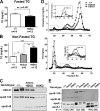
FIGURE 1.
FIGURE 1.
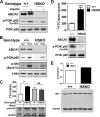
Targeted deletion of hepatocyte ABCA1 induces hypertriglyceridemia. Plasma was collected from fasted (A) and non-fasted (B–E) wild type (+/+), heterozygous (Hetero), and homozygous HSKO mice. A, fasted (6 h) plasma TG concentrations, mean ± S.E. ns, not significant. B, non-fasted plasma TG concentrations, mean ± S.E. *, p < 0.05; **, p < 0.01; ***, p < 0.001. C, Western blot analysis of hepatic ABCA1 expression and plasma levels of apoB100 and apoB48. Twenty μl of plasma were immunoprecipitated with anti-human apoB antibody, and the immunoprecipitates were fractionated by SDS-PAGE and Western blotted using the same anti-human apoB antibody. D, FPLC profiles of cholesterol (top) and triglyceride (bottom) in pooled plasma (450 μl; n = 6/genotype). Insets show the VLDL cholesterol (top) and LDL TG (bottom) results. E, Western blot analysis of 25 μl of selected FPLC fractions corresponding to D.
FIGURE 2.
FIGURE 2.
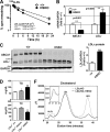
Selective deletion of hepatocyte ABCA1 increases hepatic TG secretion and reduces post-heparin lipase activity. A, TG mass accumulation per g of liver during isolated recirculating liver perfusion of WT (+/+) (n = 6) and HSKO (n = 5) mice. Data points denote mean TG ± S.E. of mice of the indicated genotype at each time point; the line of best fit, determined by linear regression analysis, is shown for both genotypes. Accumulation rates (μg/g liver/min) of TG were derived by linear regression analysis of the slope of the plot for each mouse. The mean ± S.E. accumulation rate was 1.87 ± 0.07 and 3.40 ± 0.20 μg/min/g liver for WT and HSKO mice, respectively. B, appearance of newly synthesized plasma TG and apoB after detergent block (n = 3 for each genotype). Data points denote the mean ± S.E. of radiolabeled TG in 100 μl of plasma; the line of best fit, determined by linear regression analysis, is shown for both genotypes. Accumulation rates were derived by linear regression analysis of the slope of the plot for each mouse; mean ± S.E. accumulation rates ([3H]TG/100 μl of plasma/h) of TG were 51,550 ± 6,205 and 136,500 ± 12,480 for WT and HSKO mice, respectively. Accumulation of newly synthesized and secreted plasma apoB in the terminal plasma sample was determined after immunoprecipitation with anti-apoB antiserum, SDS-PAGE separation of proteins, and PhosphorImager analysis. Relative PhosphorImager intensity of total apoB (apoB100 + B48), normalized to a WT mouse sample, is shown below the gel. C, plasma isolated 15 min after intravenous heparin injection (300 units/kg) was used to measure the HL and LPL activity in WT (+/+, n = 8) and HSKO (n = 7) mice. D, plasma TG and TC concentration after oral gavage of olive oil (150 μl) in WT (+/+) and HSKO mice (n = 4/group). Data expressed in mean ± S.E. *, p < 0.05; ***, p < 0.001 by Student's t test.
FIGURE 3.
FIGURE 3.
Targeted deletion of hepatocyte ABCA1 does not alter liver lipid content, gene expression, or lipid fatty acid composition. A, liver lipid content (TG, TC, free cholesterol (FC), and cholesteryl ester (CE)) in chow-fed wild type (+/+, n = 11) and HSKO (n = 9) mice was analyzed by enzymatic assays, and results were normalized to liver wet weight. B, mRNA expression of genes related to lipid metabolism were analyzed by quantitative real-time PCR and normalized to GAPDH expression. Mean ± S.E., n = 6/genotype. C, fatty acid (FA) composition of hepatic cholesteryl ester, TG, and phospholipid (PL) from wild type (+/+, n = 3) and HSKO (+/+, n = 3) mice was determined by gas-liquid chromatography. Percentage distribution of polyunsaturated fatty acids (PUFA; n-3 and n-6), monounsaturated fatty acids (MUFA), and saturated fatty acids (SFA) is shown in each column. D, hepatic free fatty acid (FFA) content was determined in liver from wild type and HSKO mice (n = 5/group) by enzymatic assay. Data are expressed as mean ± S.E. ns, not significant at p = 0.05 by Student's t test.
FIGURE 4.
FIGURE 4.
Targeted deletion of hepatocyte ABCA1 increases plasma VLDL size. A, fractionation of VLDL1 (Sf = 100–400) and VLDL2 (Sf = 20–100) in pooled plasma (n = 3/genotype, 200 μl total) by density gradient ultracentrifugation. VLDL1 and -2 were then assayed for TG concentration. B, VLDL size determination of pooled VLDL1 and VLDL2 fractions (equal volumes) by dynamic laser light scattering. C, VLDL fractions from A were concentrated, and 4 μg of VLDL protein was separated by 4–16% SDS-PAGE and transferred to polyvinylidene difluoride membrane for Western blot analysis of apoB, apoE, and apoA-I.
FIGURE 5.
FIGURE 5.
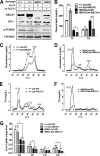
Targeted deletion of hepatocyte ABCA1 induces defective PI3K signaling. A, Western blot analysis of liver ABCA1, PI3K p85, and phospho-PI3K p85 expression 5 min after insulin (5 units/kg) injection into the portal vein. B, Western blot analysis of liver phospho-PI3K p85 (p-PI3Kp85), phospho-Akt (p-Akt), and total Akt (t-Akt) expression after fasting or fasting/refeeding of mice. C, PI3K inhibitor, wortmannin (1.5 μg/g body weight in DMSO), or DMSO alone was injected into the peritoneal cavity of mice 1 h prior to injection of detergent (1000 mg of poloxamer 407/kg of body weight) to block lipolysis and 7 μCi of [35S]Cys/Met/g of body weight as described under “Experimental Procedures.” Plasma TG concentration (mean ± S.E., n = 3) and apoB phosphor image (bottom) were analyzed 2 h after detergent block. Relative PhosphorImager intensity of apoB is shown below each lane. Values were normalized to the apoB100 band in the DMSO-treated WT mouse. D, triglyceride secretion ([3H]TG) and PI3K phosphorylation from isolated primary hepatocytes. E, quantification of MTP expression in livers of WT and HSKO mice by real-time PCR (mRNA pool from 3 mice/genotype; top) and Western blot analysis (n = 3 of each genotype; bottom). Data were normalized to GAPDH expression. Data are expressed as mean ± S.E. *, p < 0.05; ***, p < 0.001 by Student's t test. ns, not significant.
FIGURE 6.
FIGURE 6.
Hepatocyte-specific deletion of ABCA1 induces rapid LDL clearance from plasma. LDL from LDLrKO mice was isolated and radioiodinated with 125I. A, 125I-LDL tracer was injected into WT and HSKO recipient mice, and periodic blood samples were taken to quantify the amount of tracer remaining in plasma (results normalized to the 2 min time point). Mean ± S.E., n = 4. FCR values (mean ± S.E.) were calculated from individual plasma die-away curves using a biexponential curve-fitting program. ***, p < 0.001 by Student's t test. B, real-time PCR quantification of hepatic ABCA1 and LDLr mRNA levels. Data were normalized to GAPDH expression (mean ± S.E.; n = 6). C, liver protein (50 μg) from WT and HSKO mice was fractionated by SDS-PAGE and analyzed for expression of ABCA1, LDLr, and GAPDH by Western blot. Relative quantification of the blot is shown on the right. Data are expressed as mean ± S.E. D, effects of hepatic ABCA1 deficiency on non-fasting plasma TG (top) and TC (bottom) in the LDLrKO background. Shown are wild type (LDLrKO, +/+), heterozygous (LDLrKO, Hetero), and homozygous HSKO (LDLrKO, HSKO) mice (n = 4/group), mean ± S.E. F, FPLC-cholesterol profiles of pooled plasma of wild type (LDLrKO, +/+) and HSKO (LDLrKO, HSKO) mice in the LDLrKO background (n = 4/group). Inset, expanded VLDL region. *, p < 0.05; **, p < 0.01; ***, p < 0.001 by Student's t test (A and B) or analysis of variance (D).
FIGURE 7.
FIGURE 7.
Adenoviral overexpression of ABCA1 normalizes plasma TG concentrations in HSKO mice. Mice were fed an HF diet for 8 weeks before intravenous injection of either Ad-AP or Ad-ABCA1. Mice were sacrificed 3 days later to collect liver and plasma samples. A, Western blot analysis of hepatic ABCA1, LDLr, phosphorylated PI3K (p-PI3K), and total PI3K (t-PI3K) expression in wild type (WT; +/+) and HSKO mice. ABCA1 and LDLr expression level were quantified (normalized to GAPDH). Expression relative to control mice is denoted below each band. B, plasma concentrations of TG and TC in WT and HSKO mice (n = 4/group) after adenovirus injection. Data are expressed as mean ± S.E. *, p < 0.05; ***, p < 0.001 by Student's t test. C, FPLC-cholesterol profile from pooled plasma of WT mice (n = 4) after adenoviral administration. D, FPCL-cholesterol profile from pooled plasma of HSKO mice (n = 4) after adenovirus administration. E, FPLC-TG profile from pooled plasma of WT mice (n = 4) after adenovirus administration. F, FPLC-TG profile from pooled plasma of HSKO mice (n = 4) after adenovirus administration. G, hepatic content of TG, TC, FC, and cholesteryl ester (CE) in WT and HSKO mice (n = 4/group). Data are expressed as mean ± S.E. *, p < 0.05 by one-way analysis of variance with Tukey's multiple comparison.

Similar articles

Cited by

  • Macrophage 12/15 lipoxygenase expression increases plasma and hepatic lipid levels and exacerbates atherosclerosis.
    Rong S, Cao Q, Liu M, Seo J, Jia L, Boudyguina E, Gebre AK, Colvin PL, Smith TL, Murphy RC, Mishra N, Parks JS. Rong S, et al. J Lipid Res. 2012 Apr;53(4):686-95. doi: 10.1194/jlr.M022723. Epub 2012 Jan 25. J Lipid Res. 2012. PMID: 22279185 Free PMC article.
  • iPS-cell-derived microglia promote brain organoid maturation via cholesterol transfer.
    Park DS, Kozaki T, Tiwari SK, Moreira M, Khalilnezhad A, Torta F, Olivié N, Thiam CH, Liani O, Silvin A, Phoo WW, Gao L, Triebl A, Tham WK, Gonçalves L, Kong WT, Raman S, Zhang XM, Dunsmore G, Dutertre CA, Lee S, Ong JM, Balachander A, Khalilnezhad S, Lum J, Duan K, Lim ZM, Tan L, Low I, Utami KH, Yeo XY, Di Tommaso S, Dupuy JW, Varga B, Karadottir RT, Madathummal MC, Bonne I, Malleret B, Binte ZY, Wei Da N, Tan Y, Wong WJ, Zhang J, Chen J, Sobota RM, Howland SW, Ng LG, Saltel F, Castel D, Grill J, Minard V, Albani S, Chan JKY, Thion MS, Jung SY, Wenk MR, Pouladi MA, Pasqualini C, Angeli V, Cexus ONF, Ginhoux F. Park DS, et al. Nature. 2023 Nov;623(7986):397-405. doi: 10.1038/s41586-023-06713-1. Epub 2023 Nov 1. Nature. 2023. PMID: 37914940
  • Triglycerides and heart disease: still a hypothesis?
    Goldberg IJ, Eckel RH, McPherson R. Goldberg IJ, et al. Arterioscler Thromb Vasc Biol. 2011 Aug;31(8):1716-25. doi: 10.1161/ATVBAHA.111.226100. Epub 2011 Apr 28. Arterioscler Thromb Vasc Biol. 2011. PMID: 21527746 Free PMC article. Review.
  • Alternations of Lipoprotein Profiles in the Plasma as Biomarkers of Huntington's Disease.
    Chang KH, Cheng ML, Lo CJ, Fan CM, Wu YR, Chen CM. Chang KH, et al. Cells. 2023 Jan 20;12(3):385. doi: 10.3390/cells12030385. Cells. 2023. PMID: 36766727 Free PMC article.
  • XX sex chromosome complement promotes atherosclerosis in mice.
    AlSiraj Y, Chen X, Thatcher SE, Temel RE, Cai L, Blalock E, Katz W, Ali HM, Petriello M, Deng P, Morris AJ, Wang X, Lusis AJ, Arnold AP, Reue K, Thompson K, Tso P, Cassis LA. AlSiraj Y, et al. Nat Commun. 2019 Jun 14;10(1):2631. doi: 10.1038/s41467-019-10462-z. Nat Commun. 2019. PMID: 31201301 Free PMC article.

References

    1. Timmins J. M., Lee J. Y., Boudyguina E., Kluckman K. D., Brunham L. R., Mulya A., Gebre A. K., Coutinho J. M., Colvin P. L., Smith T. L., Hayden M. R., Maeda N., Parks J. S. (2005) J. Clin. Invest. 115, 1333–1342 - PMC - PubMed
    1. Brunham L. R., Kruit J. K., Iqbal J., Fievet C., Timmins J. M., Pape T. D., Coburn B. A., Bissada N., Staels B., Groen A. K., Hussain M. M., Parks J. S., Kuipers F., Hayden M. R. (2006) J. Clin. Invest. 116, 1052–1062 - PMC - PubMed
    1. Wellington C. L., Walker E. K., Suarez A., Kwok A., Bissada N., Singaraja R., Yang Y. Z., Zhang L. H., James E., Wilson J. E., Francone O., McManus B. M., Hayden M. R. (2002) Lab. Invest. 82, 273–283 - PubMed
    1. Assman G., von Eckardstein A., Brewer H. B., Jr. (2001) in The Metabolic and Molecular Bases of Inherited Disease (Scriver C. R., Beaudet A. L., Sly W. S., Valle D., Childs B., Kinzler K. W., Volkman B. F. eds) pp. 2937–2960, McGraw-Hill, New York
    1. Bodzioch M., Orsó E., Klucken J., Langmann T., Böttcher A., Diederich W., Drobnik W., Barlage S., Büchler C., Porsch-Ozcürümez M., Kaminski W. E., Hahmann H. W., Oette K., Rothe G., Aslanidis C., Lackner K. J., Schmitz G. (1999) Nat. Genet. 22, 347–351 - PubMed

Publication types

MeSH terms

LinkOut - more resources