. 2010 Apr 16;285(16):11827-35.
doi: 10.1074/jbc.M109.072744. Epub 2010 Feb 23.

Fas apoptosis inhibitory molecule regulates T cell receptor-mediated apoptosis of thymocytes by modulating Akt activation and Nur77 expression

Affiliations

Fas apoptosis inhibitory molecule regulates T cell receptor-mediated apoptosis of thymocytes by modulating Akt activation and Nur77 expression

Jianxin Huo et al. J Biol Chem. .

Abstract

Fas apoptosis inhibitory molecule (FAIM) has been demonstrated to confer resistance to Fas-induced apoptosis of lymphocytes and hepatocytes in vitro and in vivo. Here, we show that FAIM is up-regulated in thymocytes upon T cell receptor (TCR) engagement and that faim(-/-) thymocytes are highly susceptible to TCR-mediated apoptosis with increased activation of caspase-8 and -9. Furthermore, injection of anti-CD3 antibodies leads to augmented depletion of CD4(+)CD8(+) T cells in the thymus of faim(-/-) mice compared with wild-type control, suggesting that FAIM plays a role in thymocyte apoptosis. Cross-linking of the TCR on faim(-/-) thymocytes leads to an elevated protein level of the orphan nuclear receptor Nur77, which plays a role in thymocyte apoptosis. Interestingly, in the absence of FAIM, there are reduced ubiquitination and degradation of the Nur77 protein. Faim(-/-) thymocytes also exhibit a defective TCR-induced activation of Akt whose activity we now show is required for Nur77 ubiquitination. Further analyses utilizing FAIM-deficient primary thymocytes and FAIM-overexpressing DO-11.10 T cells indicate that FAIM acts upstream of Akt during TCR signaling and influences the localization of Akt to lipid rafts, hence affecting its activation. Taken together, our study defined a TCR-induced FAIM/Akt/Nur77 signaling axis that is critical for modulating the apoptosis of developing thymocytes.


Figures
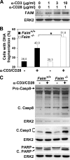
FIGURE 1.
FIGURE 1.
Enhanced TCR-mediated apoptosis of faim−/− thymocytes. A, TCR stimulation induces FAIM protein expression in thymocytes. WT thymocytes were treated with varying doses of anti-CD3/CD28 antibodies for 18 h, and cell lysates were probed for FAIM protein using specific antibodies. An anti-ERK2 blot was included as the loading control. Data shown are representatives of three independent experiments. B, graphical representation of the increase in TCR-induced apoptosis in WT and faim−/− thymocytes. Apoptosis was assessed after 18 h of stimulation with plate-bound anti-CD3 (10 μg/ml) and anti-CD28 (1 μg/ml) antibodies. Apoptosis was measured by propidium iodide staining of cellular DNA content and shown as a percentage of sub-G1 to the whole population. Numbers indicate the mean percentage of cells with DNA fragmentation ± S.D. Data shown are representative of five independent experiments. *, p = 0.024 (two-sided Student's t test). C, Western blot analyses of caspase-8 and -3 activation and poly(ADP-ribose) polymerase cleavage in WT and faim−/− thymocytes after 18 h of anti-CD3/CD28 antibodies stimulation. Data shown are representative of six independent experiments. ERK2 blots were included as the loading control. Casp8, caspase-8; PARP, poly(ADP-ribose) polymerase.
FIGURE 2.
FIGURE 2.
Augmented depletion of CD4+CD8+ thymocytes in faim−/− mice after in vivo anti-CD3 antibody treatment. A, flow cytometry analysis of thymocytes isolated from 8-week-old WT and faim−/− mice injected intraperitoneally with PBS or anti-CD3 antibody (25 μg). Percentages of cells in various thymic populations were shown. B, enumeration of cells in the various thymic fractions of faim+/+ and faim−/− mice after intraperitoneal injection with anti-CD3 antibody. *, p = 0.036 (two-sided Student's t test). DN, double negative; DP, double positive. C, increased level of FAIM protein expression in WT thymocytes after anti-CD3 antibody injection in vivo. Cell lysates were prepared from thymocytes of mice injected 16 or 48 h earlier with anti-CD3 antibody and probed with anti-FAIM or anti-ERK2 antibody. D, WT and faim−/− mice were intraperitoneally injected with anti-CD3 antibody (25 μg) 16 h earlier, and whole cell lysates of thymocytes were probed for activated caspase-8 (Casp8) and -3 (Casp3).
FIGURE 3.
FIGURE 3.
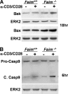
FAIM deficiency leads to elevated levels of Bak and Bax proteins and increased activation of caspase-9 during TCR-mediated apoptosis of thymocytes. A, increased protein expression of Bak and Bax in faim−/− thymocytes. WT and faim/ thymocytes were stimulated with anti-CD3 (10 μg/ml) and anti-CD28 (1 μg/ml) antibodies for 18 h and analyzed for Bax and Bak induction via Western blot analysis. B, enhanced caspase-9 (Casp9) activation in faim−/− thymocytes. Thymocytes from WT and faim−/− mice were treated with anti-CD3/CD28 antibodies for 6 h, and cell lysates were probed for increase in the activated form of caspase-9. Anti-ERK2 blots were included as loading controls. Data are representative of four independent experiments.
FIGURE 4.
FIGURE 4.
FAIM regulates Nur77 protein expression in TCR-stimulated thymocytes. A, shown is a real time reverse transcription-PCR analysis of Nur77 mRNA expression in WT and faim−/− thymocytes stimulated with anti-CD3/CD28 antibodies for 6 h. TaqMan assays for Nur77 and glyceraldehyde-3-phosphate dehydrogenase were purchased from Applied Biosystems. Data are representative of three independent experiments. B, Nur77 protein levels in faim−/− thymocytes were increased after TCR stimulation. Western blot analysis of Nur77 protein expression in faim+/+ and faim−/− thymocytes at 6- and 18-h time points after stimulation with anti-CD3 (10 μg/ml) and anti-CD28 (1 μg/ml) antibodies. An ERK2 blot was included as loading control. Data are representative of five independent experiments. C, shown is a reduced induction of Nur77 protein expression in DO-11.10 cells overexpressing FAIM. Cells were transfected with pcDNA3.1-FLAG (flag) or pcDNA3.1-FLAG-FAIM (flag-FAIM) plasmid and subsequently stimulated with anti-CD3/CD28 antibodies for 18 h and analyzed via Western blot for Nur77 protein levels. The FAIM and ERK2 blots were included as control for transfection and loading, respectively. Data are representative of three independent experiments. D, overexpression of FAIM reduces TCR-mediated apoptosis in DO-11.10 cells. Graphical representation of the percentage of cells with DNA fragmentation of TCR-stimulated DO-11.10 cells transfected with pcDNA3.1-FLAG (flag) or pcDNA3.1-FLAG-FAIM (flag-FAIM) plasmid. Calculation of apoptosis is as defined in Fig. 1. Numbers indicate mean percentage of cells with DNA fragmentation ± S.E. (n = 4); *, p = 0.010 (two-sided Student's t test). E, FAIM promotes Nur77 degradation after TCR stimulation. Western blot analysis of Nur77 protein expression in faim+/+ and faim−/− thymocytes stimulated with anti-CD3 (10 μg/ml) and anti-CD28 (1 μg/ml) antibodies for 18 h. The protein synthesis inhibitor cycloheximide (CHX) (1 μg/ml) was added in the last hour of stimulation. An ERK2 blot was included as loading control. Data are representative of three independent experiments. F, FAIM affects Nur77 ubiquitination in TCR-stimulated thymocytes. faim+/+ and faim−/− thymocytes were stimulated with anti-CD3 (10 μg/ml) and anti-CD28 (1 μg/ml) antibodies for 18 h. Nur77 protein was immunoprecipitated (IP) with an anti-Nur77 monoclonal antibody and subsequently probed for ubiquitination. Data shown are representative of four independent experiments. KO, knock-out; IB, immunoblot.
FIGURE 5.
FIGURE 5.
Akt activation is regulated by FAIM and affects Nur77 protein levels. A, reduced Akt activation in faim−/− thymocytes. Western blot analysis of Akt threonine 308 phosphorylation in WT and faim−/− thymocytes stimulated via the TCR. Data shown are representative of three independent experiments. B, overexpression of FAIM leads to the sustained activation of Akt during TCR engagement. DO-11.10 cells were transfected with pcDNA3.1-FLAG (flag) or pcDNA3.1-FLAG-FAIM (flag-FAIM) plasmid and examined for Akt phosphorylation after anti-CD3/CD28 stimulation by Western blot analysis. The anti-Akt1, anti-FAIM, anti-FLAG, and anti-ERK2 blots served as controls for transfections and loading of cell lysates, respectively. Data shown are representative of three independent experiments. C and D, Akt controls the protein level of Nur77. DO-11.10 cells (C) or WT thymocytes (D) were stimulated with anti-CD3/CD28 antibodies for 18 h in the absence or presence of varying concentrations of the Akt inhibitor (1,3-dihydro-1-(1-((4-(6-phenyl-1H-imidazo[4,5-g]quinoxalin-7-yl)phenyl)methyl)-4-piperidinyl)-2H-benzimidazol-2-one trifluoroacetate salt) and examined for the protein levels of Nur77 in Western blot analysis. Anti-ERK2 blot was included as loading controls. Data shown are representative of three independent experiments. E, Akt regulates Nur77 ubiquitination in TCR-stimulated thymocytes. WT thymocytes were stimulated with anti-CD3 (10 μg/ml) and anti-CD28 (1 μg/ml) antibodies for 18 h with varying concentrations of the Akt inhibitor (Akt inhi.). Nur77 protein was immunoprecipitated (IP) with an anti-Nur77 monoclonal antibody and probed for Nur77 ubiquitination. Data shown are representative of three independent experiments. IB, immunoblot.
FIGURE 6.
FIGURE 6.
FAIM facilitates the recruitment of Akt into lipid rafts. A, FAIM and Akt localize to lipid rafts upon TCR stimulation. WT thymocytes were nontreated (−) or stimulated (+) via the TCR for 30 min and fractionated and examined for the presence of Akt1 and FAIM proteins. Fraction 4 represents the lipid raft fraction as indicated by the presence of lipid raft-located linker for activation of T cells. Data are representative of three independent fractionation experiments. B, shown are reduced Akt in lipid rafts of faim−/− thymocytes upon TCR stimulation. Cells were treated and processed as above to examine the presence of Akt1 and FAIM in lipid rafts. Only fractions 3 and 4, which represented the lipid raft fractions, are shown. Data are representative of three independent experiments. An anti-linker for activation of T cells (LAT) blot was included to indicate the lipid raft fractions. C, shown is an increased amount of Akt in lipid rafts of FAIM-overexpressing DO-11.10 cells upon TCR stimulation. Cells were transfected with pcDNA3.1-FLAG (flag) or pcDNA3.1-FLAG-FAIM (flag-FAIM) plasmid and stimulated with anti-CD3/CD28 antibodies for 30 min and subsequently fractionated and examined as above for the presence of Akt1, FLAG-FAIM, and endogenous FAIM in lipid raft factions. Only fractions 3 and 4 representing the raft fractions are shown. Data shown are representative of three independent experiments.

Similar articles

Cited by

References

    1. Rathmell J. C., Thompson C. B. (2002) Cell 109, S97–107 - PubMed
    1. Sohn S. J., Rajpal A., Winoto A. (2003) Curr. Opin. Immunol. 15, 209–216 - PubMed
    1. Dhein J., Walczak H., Bäumler C., Debatin K. M., Krammer P. H. (1995) Nature 373, 438–441 - PubMed
    1. Moulian N., Berrih-Aknin S. (1998) Semin. Immunol. 10, 449–456 - PubMed
    1. Werlen G., Hausmann B., Naeher D., Palmer E. (2003) Science 299, 1859–1863 - PubMed

Publication types

MeSH terms