

Chronic active herpes simplex type 2 encephalitis in an asymptomatic immunocompetent child
- PMID: 20179002
- PMCID: PMC3376078
- DOI: 10.1177/0883073809353449
Chronic active herpes simplex type 2 encephalitis in an asymptomatic immunocompetent child
Erratum in
- J Child Neurol. 2010 Dec;25(12):1599
Abstract
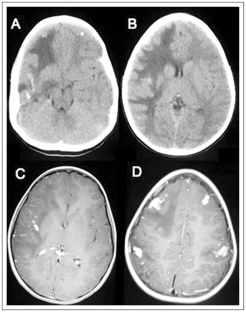
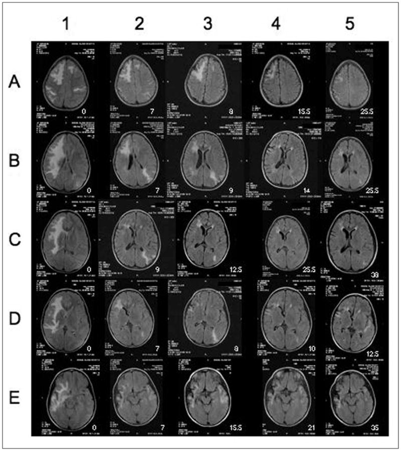
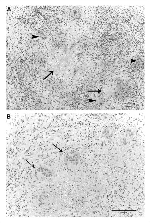
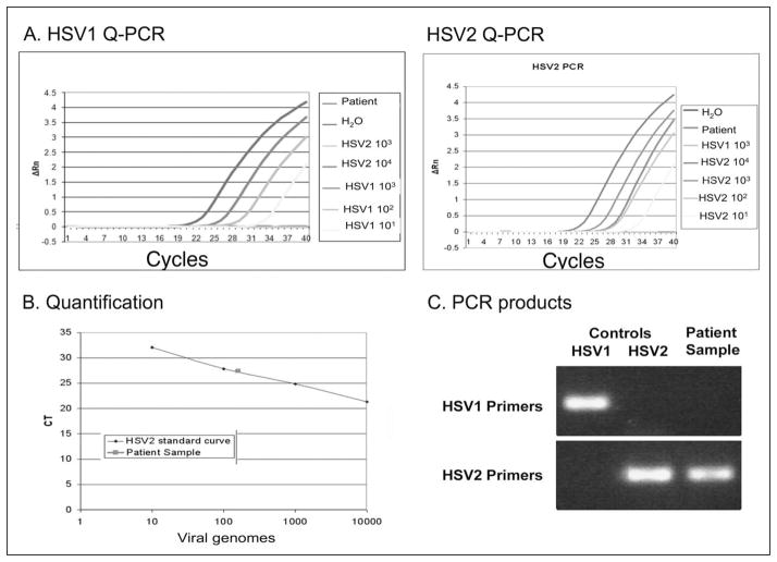
A unique form of chronic, active, granulomatous herpes simplex type 2 encephalitis is described in an asymptomatic, immunocompetent 8-year-old girl who acquired the virus as a neonate. The extensive, bilateral cerebral parenchymal involvement was discovered incidentally. Diagnosis was confirmed by a combination of serial neuroimaging, brain biopsy, and quantitative polymerase chain reaction targeted to DNA sequences in the glycoprotein G gene, allowing differentiation between herpes simplex virus types 1 and 2. The clinical course over a 5-year period, treatment with intermittent intravenous steroids, and daily valacyclovir, diagnostic imaging, and laboratory studies are reviewed in detail. This form of herpes simplex virus type 2 encephalitis hasn't been described previously and is significant because of its prolonged indolent course, absence of neurological findings or suggestive history, and benign behavior in this child, who is now 14 years old. The authors believe this entity can be unsuspected and underdiagnosed in the general pediatric population, especially in those with a prior maternal history of herpes simplex virus type 2 infection.
Conflict of interest statement
The authors declared no conflicts of interest with respect to the authorship and/or publication of this article.
Figures
Similar articles
-
Discrepant findings between proton magnetic resonance spectroscopy and magnetic resonance imaging in an immunocompetent patient with herpes simplex virus type 1 encephalitis.Scand J Infect Dis. 2012 Apr;44(4):315-9. doi: 10.3109/00365548.2011.633551. Epub 2011 Nov 28. Scand J Infect Dis. 2012. PMID: 22122735
-
Granulomatous herpes simplex encephalitis in an infant with multicystic encephalopathy: a distinct clinicopathologic entity?Pediatr Neurol. 2014 Apr;50(4):392-6. doi: 10.1016/j.pediatrneurol.2013.12.008. Epub 2013 Dec 14. Pediatr Neurol. 2014. PMID: 24485930
-
Atypical herpes type 2 encephalitis associated with normal MRI imaging.J Neurol Neurosurg Psychiatry. 2003 Jul;74(7):974-6. doi: 10.1136/jnnp.74.7.974. J Neurol Neurosurg Psychiatry. 2003. PMID: 12810797 Free PMC article.
-
[Difficulties in early diagnosis of Herpes simplex encephalitis].Pol Merkur Lekarski. 2005 Nov;19(113):719-22. Pol Merkur Lekarski. 2005. PMID: 16498820 Review. Polish.
-
Herpes Simplex Type 2 Encephalitis After Craniotomy: Case Report and Literature Review.World Neurosurg. 2016 Apr;88:691.e9-691.e12. doi: 10.1016/j.wneu.2015.11.101. Epub 2015 Dec 25. World Neurosurg. 2016. PMID: 26724620 Review.
Cited by
-
Chronic herpes simplex type-1 encephalitis with intractable epilepsy in an immunosuppressed patient.Infection. 2016 Feb;44(1):121-5. doi: 10.1007/s15010-015-0822-6. Epub 2015 Jul 18. Infection. 2016. PMID: 26187268
-
The case for immunomodulatory approaches in treating HSV encephalitis.Future Virol. 2013 Mar 1;8(3):259-272. doi: 10.2217/fvl.12.138. Future Virol. 2013. PMID: 23956785 Free PMC article.
-
Herpes simplex virus dances with amyloid precursor protein while exiting the cell.PLoS One. 2011 Mar 31;6(3):e17966. doi: 10.1371/journal.pone.0017966. PLoS One. 2011. PMID: 21483850 Free PMC article.
-
Herpes Simplex Virus 1 and 2 Infections during Differentiation of Human Cortical Neurons.Viruses. 2021 Oct 14;13(10):2072. doi: 10.3390/v13102072. Viruses. 2021. PMID: 34696502 Free PMC article.
-
Herpes Simplex Virus, Alzheimer's Disease and a Possible Role for Rab GTPases.Front Cell Dev Biol. 2019 Aug 7;7:134. doi: 10.3389/fcell.2019.00134. eCollection 2019. Front Cell Dev Biol. 2019. PMID: 31448273 Free PMC article.
References
-
- Love S, Koch P, Urbach H, Dawson TP. Chronic granulomatous herpes simplex encephalitis in children. J Neuropathol Exp Neurol. 2004;63:1173–1181. - PubMed
-
- Preiser W, Weber B, Klös G, et al. Unusual course of herpes simplex virus encephalitis after acyclovir therapy. Infection. 1996;24:384–389. - PubMed
-
- Hori T, Suzuki T, Terashima Y, et al. Chronic herpes simplex encephalitis with somnambulism: CT, MR, and SPECT findings. Jpn J Psychiatry Neurol. 1990;44:735–739. - PubMed
-
- Jay V, Blaser S, Hwang P, et al. Pathology of chronic herpes infection associated with seizure disorder: a report of two cases with tissue detection of herpes simplex virus 1 by the polymerase chain reaction. Ped Pathol Lab Med. 1995;15:131–146. - PubMed
-
- Assenbauer B, McEntagart M, King MD, et al. Chronic active destructive herpes simplex encephalitis with recovery of viral DNA 12 years after disease onset. Neuropediatrics. 1998;29:120–123. - PubMed
Publication types
MeSH terms
Grants and funding
LinkOut - more resources
Full Text Sources
