Review
. 2010 Feb;22(2):296-306.
doi: 10.1105/tpc.109.072777. Epub 2010 Feb 23.

Out of the mouths of plants: the molecular basis of the evolution and diversity of stomatal development

Affiliations
Review

Out of the mouths of plants: the molecular basis of the evolution and diversity of stomatal development

Kylee M Peterson et al. Plant Cell. 2010 Feb.

Abstract

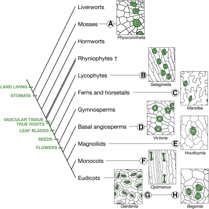
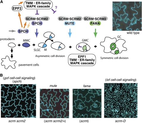
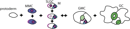
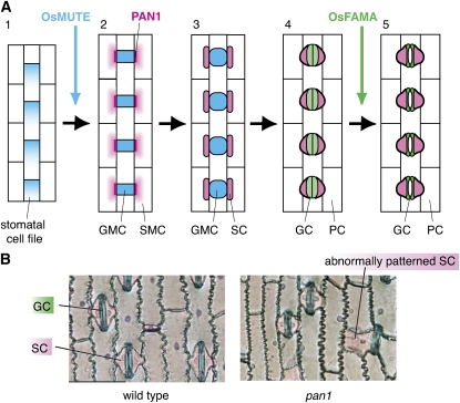
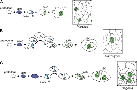
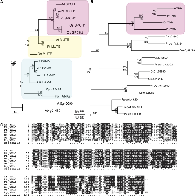
Stomata are microscopic valves on the plant epidermis that played a critical role in the evolution of land plants. Studies in the model dicot Arabidopsis thaliana have identified key transcription factors and signaling pathways controlling stomatal patterning and differentiation. Three paralogous Arabidopsis basic helix-loop-helix proteins, SPEECHLESS (SPCH), MUTE, and FAMA, mediate sequential steps of cell-state transitions together with their heterodimeric partners SCREAM (SCRM) and SCRM2. Cell-cell signaling components, including putative ligands, putative receptors, and mitogen-activated protein kinase cascades, orient asymmetric cell divisions and prevent overproduction and clustering of stomata. The recent availability of genome sequence and reverse genetics tools for model monocots and basal land plants allows for the examination of the conservation of genes important in stomatal patterning and differentiation. Studies in grasses have revealed that divergence of SPCH-MUTE-FAMA predates the evolutionary split of monocots and dicots and that these proteins show conserved and novel roles in stomatal differentiation. By contrast, specific asymmetric cell divisions in Arabidopsis and grasses require unique molecular components. Molecular phylogenetic analysis implies potential conservation of signaling pathways and prototypical functions of the transcription factors specifying stomatal differentiation.


Figures

Figure 1.
Figure 1.
Diversity of Stomata across Land Plant Taxa. A phylogenetic tree of extant and extinct (†) land plants includes evolutionary traits supporting success on land. The wide diversity of stomatal complexes among these groups is represented by epidermal tracings of P. patens ([A]; nonvascular), Selaginella kraussiana ([B]; lycophyte, vascular), Marsilea macropoda ([C]; fern, vascular), Victoria amazonica ([D]; Nymphaeaceae, basal angiosperm), Houttuynia cordata ([E]; Piperales, magnoliid), Oplimenus hirtellus ([F]; Poales, monocot grass), Gardenia taitensis ([G]; Gentianales, eudicot angiosperm), and Begonia rex-cultorumRoberta’ ([H]; Cucurbitales, eudicot angiosperm). Stomata are colored green. Note that the stomata of Physcomitrella have a single GC, while the GCs of Oplimenus, a grass, have a dumbbell shape. Only Houttuynia, Gardenia, and Begonia show evidence of asymmetric amplifying divisions within the stomatal lineage. Physcomitrella is traced from a scanning electron microscopy image by L. Pillitteri.
Figure 2.
Figure 2.
Stomatal Development in Arabidopsis. (A) Schematic diagram of stomatal development. The cell states of stomatal precursors are driven by three paralogous bHLH transcription factors, which likely dimerize with SCRM and SCRM2 as a mechanism for coordinated action. Initial specification of the stomatal cell lineage, in which a protodermal cell becomes a meristemoid mother cell (MMC), is controlled by SPCH. Protodermal cells not entering the stomatal lineage differentiate into pavement cells. The MMC divides asymmetrically to form a meristemoid (M) and SLGC and may reiterate similar divisions several times. MUTE controls the cell-state transition from M to GMC, and FAMA is required for correct division of the GMC into GCs forming a functional stoma. It is proposed that a MAP kinase signaling cascade following putative ligands EPF1 and EPF2 (EPF1 expressed in GMC, light green, and EPF2 expressed in MMC, blue, and M, cyan) perceived by TMM and the ER family of RLKs acts to suppress stomatal identity in cells adjacent to developing stomata; new meristemoids can differentiate at least one cell away, as shown near GMC. An image of wild-type epidermis is shown at the top right. (B) Epidermal phenotypes of stomatal differentiation mutants. Shown are the rosette leaf epidermis of (from left) scrm scrm2, mute, fama, and scrm-D. scrm scrm2 produces epidermis solely composed of pavement cells, a phenotype identical to that of spch as well as gain-of-function mutants in stomatal cell–cell signaling genes. mute and fama produce epidermis with arrested stomatal precursor cells similar to scrm scrm2/+ and scrm, respectively. scrm-D produces epidermis solely composed of stomata, a phenotype similar to loss of function in stomatal signaling genes. Images are reproduced from Kanaoka et al. (2008).
Figure 3.
Figure 3.
Localization of BASL. BASL acts as a molecular signal instructing stomatal lineage cells to divide away from it. BASL protein appears initially in the nuclei of meristemoid mother cells (MMC), which differentiate from protodermal cells. The protein then localizes in a second location at the cell periphery opposite the site of the future asymmetric division. Following that division, BASL remains at the cell periphery but fades away from the nucleus of the larger daughter cell (SLGC), which loses stomatal lineage identity; it remains in the nucleus of the meristemoid (M), which may further asymmetrically divide. BASL is not found in later stomatal lineage cells, such as (GMCs or GCs. However, when satellite meristemoids are formed by SLGCs that resume stomatal lineage fate, BASL appears at the SLGC periphery next to the stomatal lineage cell, providing a mechanism for maintenance of the one-cell spacing rule. (Based on data presented in Dong et al. [2009].)
Figure 4.
Figure 4.
Stomatal Development in Grasses. (A) Schematic diagram of stomatal development in grasses. (1) During early epidermal development in grasses, stomatal and nonstomatal cell files are specified, and cell division polarity is established in the stomatal cell file. This polarity will ensure that the one-cell spacing rule is maintained. (2) Os MUTE controls a single asymmetric division toward the leaf apex in the stomatal cell file, which creates GMCs (blue). Neighboring cell files (SMC; pink gradient) receive a signal via putative receptor PAN1 (magenta), which localizes at the area of GMC contact and polarize in preparation for division. (3) SMCs divide asymmetrically toward PAN1 to form SCs (pink), which will act as ion reservoirs for the operation of mature stomata. (4) GMCs divide once symmetrically to form GCs (light green). (5) Finally, GC and SC terminally differentiate, forming mature dumbbell-shaped stomata (dark green). Os FAMA is required for the differentiation of GCs, though not their symmetric division. (B) Leaf epidermal peel from the wild type (left) and pan1 mutant maize (right). GC and SC are stained in blue and pink, respectively. Unlike the wild type, the pan1 mutant occasionally fails to produce proper asymmetric divisions that give rise to SC, resulting in abnormal SC patterning. Images kindly provided by Laurie Smith (University of California, San Diego).
Figure 5.
Figure 5.
Developmental Hypotheses for Stomatal Complex Diversity. (A) Stomata in the fern Marsilea appear to develop through a process lacking amplifying divisions of a meristemoid (cf. Arabidopsis in Figure 2A). (B) Houttuynia (magnoliid) stomata are surrounded by a spiral arrangement of cells that suggests a large number of amplifying divisions. (C) In Begonia, a eudicot, stomata arise in groups that can be explained by early division of a stomatal precursor, such as an MMC, and retention of MMC identity by the daughter cells.
Figure 6.
Figure 6.
Stomatal Patterning and Spacing Genes Are Conserved among Embryophytes. (A) A gene tree of master regulatory bHLH transcription factors (SPCH, MUTE, and FAMA) in stomatal development. Amino acid sequences from bHLH and ACT domains were aligned and the tree constructed using Bayesian and neighbor-joining methods. The Bayesian phylogram is shown. The pink, yellow, and cyan rectangles highlight the SPCH, MUTE, and FAMA clades, respectively. Posterior probability values are indicated above the nodes, and bootstrap values over 50 (100,000 replicates) are indicated below the nodes. (B) A gene tree of TMM, which encodes an LRR receptor-like protein necessary for proper stomatal spacing in Arabidopsis. The entire amino acid sequence was aligned and the tree constructed as in (A). The Bayesian phylogram is shown. The pink rectangle highlights the single copy TMM clade. Posterior probability values are indicated above the nodes, and bootstrap values over 50 (100,000 replicates) are indicated below the nodes. (C) Amino acid alignment of the N-terminal portion of YODA (YDA), a MAPKKK, which is essential to regulate the activity of the protein in Arabidopsis. YDA is also required for appropriate stomatal spacing. At, Arabidopsis thaliana; Pt, Populus trichocarpa; Os, Oryza sativa; Pp, Physcomitrella patens. See Supplemental Methods and Supplemental Table 1 online for detailed bioinformatic and phylogenetic methods and gene ID numbers.

Similar articles

Cited by

References

    1. Abrash E.B., Bergmann D.C. (2010). Regional specification of stomatal production by the putative ligand CHALLAH. Development 137: 447–455 - PubMed
    1. Apostolakos P., Panteris E., Galatis B. (1997). Microtubule and actin filament organization during stomatal morphogenesis in the fern Asplenium nidus. 1. Guard cell mother cell. Protoplasma 198: 93–106 - PubMed
    1. Berger D., Altmann T. (2000). A subtilisin-like serine protease involved in the regulation of stomatal density and distribution in Arabidopsis thaliana. Genes Dev. 14: 1119–1131 - PMC - PubMed
    1. Bergmann D.C., Lukowitz W., Somerville C.R. (2004). Stomatal development and pattern controlled by a MAPKK kinase. Science 304: 1494–1497 - PubMed
    1. Bhave N.S., Veley K.M., Nadeau J.A., Lucas J.R., Bhave S.L., Sack F.D. (2009). TOO MANY MOUTHS promotes cell fate progression in stomatal development of Arabidopsis stems. Planta 229: 357–367 - PubMed

Publication types

MeSH terms

LinkOut - more resources