. 2010 Apr 22;115(16):3320-8.
doi: 10.1182/blood-2009-02-202440. Epub 2010 Feb 23.

Rac1 targeting suppresses p53 deficiency-mediated lymphomagenesis

Affiliations

Rac1 targeting suppresses p53 deficiency-mediated lymphomagenesis

Emily E Bosco et al. Blood. .

Abstract

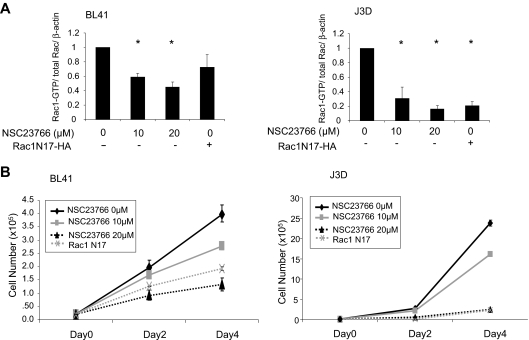
Mutation of the p53 tumor suppressor is associated with disease progression, therapeutic resistance, and poor prognosis in patients with lymphoid malignancies and can occur in approximately 50% of Burkitt lymphomas. Thus, new therapies are needed to specifically target p53-deficient lymphomas with increased efficacy. In the current study, the specific impact of inhibition of the small GTPase Rac1 on p53-deficient B- and T-lymphoma cells was investigated. p53 deficiency resulted in increased Rac1 activity in both B-cell and T-cell lines, and its suppression was able to abrogate p53 deficiency-mediated lymphoma cell proliferation. Further, Rac targeting resulted in increased apoptosis via a p53-independent mechanism. By probing multiple signaling axes and performing rescue studies, we show that the antiproliferative effect of Rac1 targeting in lymphoma cells may involve the PAK and Akt signaling pathway, but not the mitogen-activated protein (MAP) kinase pathway. The effects of inhibition of Rac1 were extended in vivo where Rac1 targeting was able to specifically impair p53-deficient lymphoma cell growth in mouse xenografts and postpone lymphomagenesis onset in murine transplantation models. Because the Rac1 signaling axis is a critical determinant of apoptosis and tumorigenesis, it may represent an important basis for therapy in the treatment of p53-deficient lymphomas.


Figures

Figure 1
Figure 1
p53 deficiency causes elevated Rac1 activity. BL41 and BL41ts cells (left panel) and J3D and J3Dts cells (right panel) were cultured for 24 hours at 32°C or 37°C. Cell lysates were subjected to GST-PAK pull-down assay and processed for Western immunoblot with anti-p53 or anti-Rac antibodies. β-actin served as a loading control. The J3D bands displayed were part of the same gel, yet were separated by other lanes. The bands from at least 2 independent experiments were quantified by densitometry and displayed below. Error bars represent SD.
Figure 2
Figure 2
Rac1 activity contributes to p53-deficient lymphoma cell hyperproliferation. (A) Cell lysates were subjected to GST-PAK pull-down assay and processed for Western immunoblot to determine Rac1 activity in BL41 (left panel) and J3D (right panel) cells after 4-day treatment with NSC23766 or transduction with a Rac1 N17 mutation. Anti-HA or anti-Rac antibodies were used and β-actin served as a loading control. Immunoblot bands from at least 3 independent experiments were quantified by densitometry and displayed. *P < .05, significant with respect to untreated control. (B) BL41 (left panel) or J3D (right panel) cells were treated with NSC23766 or transduced with Rac1N17 as in panel A and cell number was monitored by trypan blue exclusion. Error bars represent SD.
Figure 3
Figure 3
Rac1 targeting modulates apoptotic response of p53-deficient lymphoma cells. BL41 or J3D cells treated with NSC23766 or transduced with Rac1N17 were harvested for flow cytometry to determine the rate of apoptosis by staining with Annexin V and 7AAD. Error bars represent SD.
Figure 4
Figure 4
Rac1 knockdown induces apoptosis in human lymphoma cells. (A) BL41 cells were transduced with either a YFP-tagged shRac1 construct or scrambled sh control (shSCR) and sorted 48 hours after infection. Cells were harvested 24 hours after sorting and immunoblotted with antibodies to Rac1, Rac2, pan-Rac, pPAK1, cleaved caspase-3, cytochrome c, and Gapdh. (B) Cells from panel A were analyzed by flow cytometry to determine the Annexin V/ 7AAD-positive population. Error bars represent SD.
Figure 5
Figure 5
Effects of Rac1 targeting on cell cycle and apoptosis may involve the PAK and Akt pathway. (A) BL41cells containing the p53ts mutant were treated with the Rac1 inhibitor, NSC23766, or transduced with a Rac1N17 mutant and immunoblotted for phospho-PAK1 and phospho-Akt (left panel), and potential effectors of the MAP kinase module (ie, ERK, JNK, p38; right panel) and their respective total protein levels. Both left and right panels were from the same immunoblot, and β-actin served as a loading control for both panels. Immunoblot bands were quantified by densitometry, and untreated controls were set to 1. (B) BL41 cells were transduced with lentivirus encoding either a YFP-tagged shRac1 construct or scrambled sh control (shSCR) in conjunction with retrovirus encoding either GFP-tagged caPAK, GFP-tagged caAkt, GFP-tagged Akt-KD, or GFP alone. Cells were harvested for flow cytometry 48 hours after infection to determine the rate of apoptosis in the YFP/GFP-copositive population by staining with Annexin V and 7AAD. (C) BL41 human B-lymphoma p53 mutant temperature-sensitive cells were incubated at either 32°C (p53 wild-type) or 37°C (p53 mutant) for 24 hours before a 3-hour pulse with 20μM wortmannin or 50μM LY294002. Cells were then harvested and subjected to GST-Pak pull-down assay and immunoblotted on the same day for Rac1, pAkt Ser473, total Akt, and Gapdh. The data are representative of 6 different experiments and error bars represent SD.
Figure 6
Figure 6
Rac1 targeting impairs p53-deficient tumor growth in vivo. (A) BL41 cells were transduced with either a YFP-tagged shRac1 construct or scrambled sh control (shSCR) and sorted as in Figure 4A. Cells were harvested 24 hours after sorting and injected (2 × 10⁶) contralaterally into the flanks of NOG mice and tumor growth was monitored. (B) Tumors from panel A were excised and weighed at day 73. Error bars represent SD.
Figure 7
Figure 7
Rac1-deficiency delays p53-deficient lymphoma growth in vivo. (A) Total bone marrow from a p53−/−MxRac1 mouse was isolated and injected (500 000 cells) by tail vein into lethally irradiated (1175 cGy, split-dose) BOYJ recipients. Blood chimerism was assessed 1 month after transplantation and mice containing approximately 90% or greater donor cells received 3 × 300 μg of intraperitoneal PolyIC injections every other day for 4 injections to delete the Rac1 gene in hematopoietic cells in vivo or were mock-injected. Animals were bled monthly to monitor genetic deletion of Rac1 by PCR. Representative PCRs of blood from the same animals 1 month (left panel) or 5 months (right panel) after PolyIC injection are displayed. (B) Survival of animals from panel A are displayed with transgenic p53−/− and p53−/−Rac2−/− animals included. (C) Representative tumors were excised from animals that underwent transplantation from panel A upon death and processed for PCR to assess levels of Rac1 DNA. (D) Tumors from panel C were snap-frozen in liquid nitrogen, lysed, and immunoblotted to determine the expression level of Rac1 protein.

Similar articles

Cited by

References

    1. Jaffe ES, Diebold J, Harris NL, Muller-Hermelink HK, Flandrin G, Vardiman JW. Burkitt's lymphoma: a single disease with multiple variants: the World Health Organization classification of neoplastic diseases of the hematopoietic and lymphoid tissues. Blood. 1999;93:1124. - PubMed
    1. Harris NL, Horning SJ. Burkitt's lymphoma–the message from microarrays. N Engl J Med. 2006;354:2495–2498. - PubMed
    1. Adams JM. Oncogene activation by fusion of chromosomes in leukaemia. Nature. 1985;315:541–542. - PubMed
    1. Bhatia K, Gutierrez M, Magrath IT. Burkitt's lymphoma cells frequently carry monoallelic DJ rearrangements. Curr Top Microbiol Immunol. 1992;182:319–324. - PubMed
    1. Capoulade C, Bressac-de Paillerets B, Lefrere I, et al. Overexpression of MDM2, due to enhanced translation, results in inactivation of wild-type p53 in Burkitt's lymphoma cells. Oncogene. 1998;16:1603–1610. - PubMed

Publication types

Substances