Review
. 2010 Mar 15;70(6):2146-57.
doi: 10.1158/0008-5472.CAN-09-4355. Epub 2010 Feb 23.

Drugging the PI3 kinome: from chemical tools to drugs in the clinic

Affiliations
Review

Drugging the PI3 kinome: from chemical tools to drugs in the clinic

Paul Workman et al. Cancer Res. .

Abstract

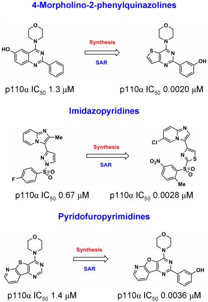
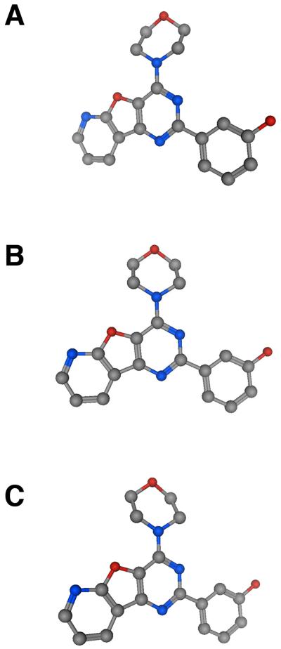
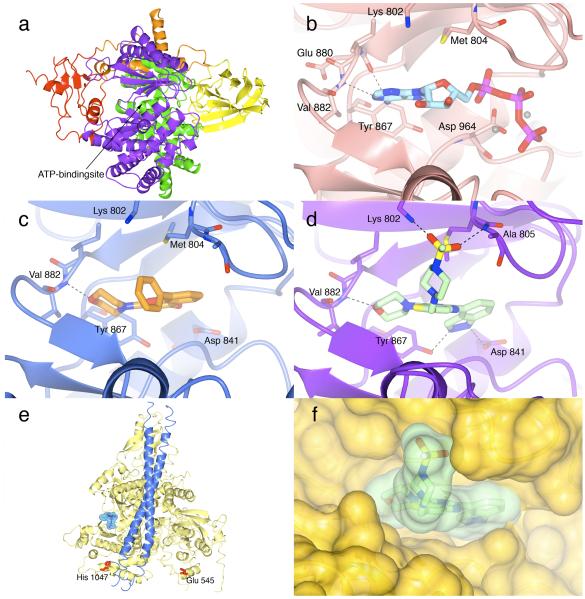
The phosphatidylinositide 3-kinase (PI3K) pathway is very commonly activated in a wide range of human cancers and is a major driving force in oncogenesis. One of the class I lipid kinase members of the PI3K family, p110alpha, is probably the most commonly mutated kinase in the human genome. Alongside genetic, molecular biological, and biochemical studies, chemical inhibitors have been extremely helpful tools in understanding the role of PI3K enzymes in signal transduction and downstream physiological and pathological processes, and also in validating PI3Ks as therapeutic targets. Although they have been valuable in the past, the early and still frequently employed inhibitors, wortmannin and LY294002, have significant limitations as chemical tools. Here, we discuss the case history of the discovery and properties of an increasingly used chemical probe, the pan-class I PI3K and mammalian target of rapamycin (mTOR) inhibitor PI-103 (a pyridofuropyrimidine), and its very recent evolution into the thienopyrimidine drug GDC-0941, which exhibits excellent oral anticancer activity in preclinical models and is now undergoing phase I clinical trials in cancer patients. We also illustrate the impact of structural biology on the design of PI3K inhibitors and on the interpretation of their effects. The challenges and outlook for drugging the PI3 kinome are discussed in the more general context of the role of structural biology and chemical biology in innovative drug discovery.


Figures

Figure 1
Figure 1
Chemical structures of the PI3K inhibitors wortmannin, LY294002, PIK-90, PIK-93, PI-103, thienopyrimidine compound 1, PI-540, PI-620 and GDC-0941. In the chemical structure of GDC-0941 the indazole moiety is highlighted in green, the sulphonylpiperazine in red and the morpholino group in blue, while the thienopyrimidine core is shown in black.
Figure 2
Figure 2
Three PI3K inhibitory hit compounds identified by high-throughput screening against p110α and their optimization into nanomolar leads. From top to bottom, the 4-morpholino-2-phenylquinazolines optimised to the thienopyrimidine compound 1, the imidazopyridines, and the pyridofurapyrimidines resulting in PI-103. For further details see references -.
Figure 3
Figure 3
Ball and stick representations of PI-103 to illustrate the planar nature of the tricyclic pyridofurapyrimidine, which limits the aqueous solubility of the compound.
Figure 4
Figure 4
a Ribbon diagram of human p110γ (PDB code 3dbs) illustrating the five-domain structure of the enzyme. The Ras-binding (RBD) domain is shown in red, the C2 domain in yellow, the helical domain in green and the bilobal catalytic domain in purple. The N-terminal ABD domain preceding the RBD-domain is shown in orange. The ATP-binding site is located in between the N- and C-terminal lobes of the catalytic domain, as indicated by the arrow. All figures were made with CCP4MG (66). b Binding of ATP to porcine p110γ (PDB code 1e8x), showing the hydrogen bond interactions of ATP with the hinge region. The protein is shown in pink, ATP is displayed in light blue. The hydrogen bond interactions with the amide group of Val 882 and with the carbonyl group of Glu 880 are shown as dotted lines. Shown in grey are the two metal ions coordinating the phosphate groups of ATP. c Binding of LY294002 to porcine p110γ (PDB code 1e7v) illustrates the crucial hydrogen bonding interaction between the oxygen of morpholino group of the weakly potent and relatively unselective inhibitor and Val 882 of the PI3K hinge region (left-hand side). The protein is shown in blue with key PI3K residues displayed and labelled. LY294002 is shown in orange. The critical hydrogen bond is shown as a dotted line. Although LY294002 extends into the affinity pocket (right-hand side) it does not fill the pocket as efficiently as the indazole moiety of GDC-0941 (see panel d). d Binding of GDC-0941 to human p110γ (PDB code 3dbs) showing the hydrogen bond interactions that anchor the inhibitor in the ATP-site. p110γ is shown in purple and GDC-0941 is shown in light green. The oxygen of the morpholino group in GDC-0941 forms a hydrogen bond with the amide of the hinge residue Val 882, (left-hand side) while the indazole moiety (right-hand side) binds deep in the affinity pocket with its two nitrogen atoms forming hydrogen bonds with the side chains of Tyr 867 and Asp 841, respectively. The oxygen atoms of the sulfonyl group (projecting out of the plane, centre) interact with the side chain of Lys 802 and the amide group of Ala 805 at the mouth of the ATP pocket. e Ribbon diagram of the p110α/p85α structure, showing p110α in yellow and the p85 niSH2 domain blue (PDB code 2rd0). To indicate the location of the ATP site, the ATP from the ATP-bound p110γ structure (PDB code 1e8x) superimposed on the p110α/p85α complex is shown. The ATP is displayed in cylinder representation with its semitransparent molecular surface superimposed in blue. Highlighted in red are the oncogenic mutation hotspots Glu 545 and His1047. f Superposition of the GDC-0941-bound p110γ structure and the human p110α/p85α structure (PDB code 2rd0). The superposition was done using the CCP4 program suite (67). p110α is shown with its solvent-accessible surface in yellow and GDC-0941 with its (semi-transparent) surface in light green. Based on this superposition, the overall fit of GDC-0941 in the p110α ATP site is very good with only a minor clash of the indazole ring with the wall of the ATP site (right-hand side). It seems likely that GDC-0941 will be able to form hydrogen bond interactions with the hinge valine (Val 851 human p110α numbering left-hand side) and with the tyrosine and aspartate side chains (Tyr 836 and Asp 810, p110α numbering; right hand side) in the affinity pocket, as observed in the GDC-0941 p110γ structure. Differences could occur in the interaction of the sulfonyl oxygens of GDC-0941 (centre) with the protein, because the equivalent of Lys 802 in p110γ is an arginine residue in p110α and the equivalent residue of Ala 805 in p110γ has a different backbone conformation in p110α.

Similar articles

Cited by

References

    1. Vanhaesebroeck B, Leevers SJ, Ahmadi K, Timms J, Katso R, Driscoll PC, Woscholski R, Parker PJ, Waterfield MD. Synthesis and function of 3-phosphorylated inositol lipids. Ann Rev Biochem. 2001;70:535–602. - PubMed
    1. Cantley LC. The phosphoinositide 3-kinase pathway. Science. 2002;296:1655–7. - PubMed
    1. Abraham RT. PI 3-kinase related kinases: ‘big’ players in stress-induced signaling pathways. DNA Repair (Amst) 2004;3:883–7. - PubMed
    1. Yuan TL, Cantley LC. PI3K pathway alterations in cancer: variations on a theme. Oncogene. 2008;27:5497–5510. - PMC - PubMed
    1. Liu P, Cheng H, Roberts TM, Zhao JJ. Disc Targeting the phosphoinositide 3-kinase pathway in cancer. Nat Rev Drug Discov. 2009;8:627–44. - PMC - PubMed

Publication types

MeSH terms