. 2010 Mar 15;70(6):2350-8.
doi: 10.1158/0008-5472.CAN-09-3885. Epub 2010 Feb 23.

Maternally expressed gene 3, an imprinted noncoding RNA gene, is associated with meningioma pathogenesis and progression

Affiliations

Maternally expressed gene 3, an imprinted noncoding RNA gene, is associated with meningioma pathogenesis and progression

Xun Zhang et al. Cancer Res. .

Abstract

Meningiomas are common tumors, representing 15% to 25% of all central nervous system tumors. NF2 gene inactivation on chromosome 22 has been shown as an early event in tumorigenesis; however, few factors underlying tumor growth and progression have been identified. The chromosomal abnormalities of 14q32 are often associated with meningioma pathogenesis and progression; therefore, it has been proposed that an as yet unidentified tumor suppressor is present at this locus. Maternally expressed gene 3 (MEG3) is an imprinted gene located at 14q32 which encodes a noncoding RNA with an antiproliferative function. We found that MEG3 mRNA is highly expressed in normal arachnoidal cells. However, MEG3 is not expressed in the majority of human meningiomas or the human meningioma cell lines IOMM-Lee and CH157-MN. There is a strong association between loss of MEG3 expression and tumor grade. Allelic loss at the MEG3 locus is also observed in meningiomas, with increasing prevalence in higher grade tumors. In addition, there is an increase in CpG methylation within the promoter and the imprinting control region of MEG3 gene in meningiomas. Functionally, MEG3 suppresses DNA synthesis in both IOMM-Lee and CH157-MN cells by approximately 60% in bromodeoxyuridine incorporation assays. Colony-forming efficiency assays show that MEG3 inhibits colony formation in CH157-MN cells by approximately 80%. Furthermore, MEG3 stimulates p53-mediated transactivation in these cell lines. Therefore, these data are consistent with the hypothesis that MEG3, which encodes a noncoding RNA, may be a tumor suppressor gene at chromosome 14q32 involved in meningioma progression via a novel mechanism.


Figures
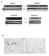
Figure 1
Figure 1
MEG3 RNA is expressed in normal human meninges but not in the majority of human meningiomas. A, RT-PCR readily detected MEG3 RNA in normal human meningeal (Lanes N), but only in 4 of 9 typical meningiomas (Top panel, left: Lane 2, 3, 4, and 5) and 1 of 11 atypical meningiomas (Top Panel, right: Lane 6). None of 7 anaplastic meningiomas (Bottom panel, left) or tumor cell lines (Bottom panel, right: Lane 1, IOMM-Lee; Lane 2, CH157-MN; Lane 3, a pituitary tumor derived cell line PDFS) expressed MEG3 RNA as examined by RT-PCR. M: molecular weight marker. B, in situ hybridization shows that MEG3 RNA is present in the arachnoidal cells of human meningeal samples (left panel), but no MEG3 RNA was detected in one typical meningioma sample (Tumor No. 5 in Fig 1A). Nuclei were stained by hematoxylin in the tumor slide.
Figure 2
Figure 2
5-aza-2′-deoxycytidine treatment results in MEG3 RNA expression in CH157-MN cells as examined by RT-PCR. M: molecular marker; C: vehicle treatment control; T: 5-aza-2′-deoxycytidine treatment.
Figure 3
Figure 3
MEG3 suppresses human meningioma cell proliferation. A, Suppression of DNA synthesis by MEG3-expression as measured by BrdU incorporation assay in IOMM-Lee cells. B, Suppression of DNA synthesis by MEG3-expressing as measured by BrdU incorporation assay in CH157-MN cells. Ctr: transfection with an empty expression vector as a control; MEG3: transfection with MEG3-expressing vector; dp: transfection with the same MEG3 expression vector but without CMV promoter; DLK: transfection with a DLK1-expressing vector. No inhibition of DNA synthesis was observed when cells were transfected with a MEG3 containing vector with the CMV promoter deleted (dp), or a DLK-expressing vector (DLK). Data are represented as mean ± SD for BrdU-labeling index from at least three independent experiments. C: In colony formation assay, transfection of MEG3- or GADD45γ-expressing vector into CH157-MN cells resulted in a significant decrease in colony number compared to the empty expression vector (Ctr), while transfection of DLK-expressing vector does not affect colony formation. D: The percentage reduction in colony numbers in each transfected cell culture plate. Data are represented as mean ± SD for BrdU-labeling index from at least three independent experiments. *: p<0.001.
Figure 4
Figure 4
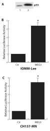
A: Both CH157-NM (Lane 1) and IOMM-Lee (Lane 2) cell line express p53 protein as examined by Western blot. B and C: MEG3 stimulates p53-mediated transactivation in IOMM-Lee (B) and CH157-NM (C) cells. The relative luciferase activity from cells without MEG3-expressing vector co-transfection is designated as 1. Data are represented as mean ± SD from at least three independent experiments. *: p<0.001.

Similar articles

Cited by

References

    1. Louis OHDN, Wiestler OD, Cavenee WK. World Health Organization Classification of Tumours of the Central Nervous System. IARC Press; Lyon: 2007.
    1. Lusis E, Gutmann DH. Meningioma: an update. Curr Opin Neurol. 2004;17(6):687–92. - PubMed
    1. Simon M, von Deimling A, Larson JJ, et al. Allelic losses on chromosomes 14, 10, and 1 in atypical and malignant meningiomas: a genetic model of meningioma progression. Cancer Res. 1995;55(20):4696–701. - PubMed
    1. Weber RG, Bostrom J, Wolter M, et al. Analysis of genomic alterations in benign, atypical, and anaplastic meningiomas: toward a genetic model of meningioma progression. Proc Natl Acad Sci U S A. 1997;94(26):14719–24. - PMC - PubMed
    1. Menon AG, Rutter JL, von Sattel JP, et al. Frequent loss of chromosome 14 in atypical and malignant meningioma: identification of a putative ‘tumor progression’ locus. Oncogene. 1997;14(5):611–6. - PubMed

Publication types

MeSH terms