. 2010 Mar 1;70(5):1814-24.
doi: 10.1158/0008-5472.CAN-09-3675. Epub 2010 Feb 23.

Vascular endothelial growth factor-C induces lymphangitic carcinomatosis, an extremely aggressive form of lung metastases

Affiliations

Vascular endothelial growth factor-C induces lymphangitic carcinomatosis, an extremely aggressive form of lung metastases

Suvendu Das et al. Cancer Res. .

Abstract

The lymphatic system is an important pathway for tumor dissemination to the lymph nodes, but to which extent it contributes to the formation of distant metastases remains unknown. We report that induction of lymphangiogenesis by vascular endothelial growth factor-C (VEGF-C) at the secondary site, in the lung, facilitates expansion of already disseminated cancer cells throughout the lung tissue. By using orthotopic spontaneous metastasis models in nude mice, we show that VEGF-C expression by tumor cells altered the pattern of pulmonary metastases from nodular to diffuse and facilitated disease progression. Metastases expressing VEGF-C were tightly associated with the airways, in contrast to the control cells that were scattered in the lung parenchyma, throughout the alveolar region. VEGF-C induced lung lymphangiogenesis and promoted intralymphatic spread of metastases in the lung and formation of tumor emboli in the pulmonary arteries. This pattern of metastasis corresponds to lymphangitic carcinomatosis metastatic phenotype in human cancer patients, an extremely aggressive pattern of pulmonary metastases. In accordance, pulmonary breast cancer metastases from patients which were classified as lymphangitic carcinomatosis showed high levels of VEGF-C expression in cancer cells. These data show that VEGF-C promotes late steps of the metastatic process and identify the VEGF-C/VEGF receptor-3 pathway as the target not only for prevention of metastases, but also for treatment of established metastatic disease.


Figures

Figure 1
Figure 1
Metastases expressing VEGF-C are associated with the airways. Fluorescence stereomicroscopy of thick lung sections (150 μm) showing distribution of MDA/pcDNA (A1, A2) and MDA/VEGF-C (B1, B2) metastases (RFP, red) in the lung. Confocal analysis of MDA/pcDNA (A3) and MDA/VEGF-C (B3) metastases. Note the tight association of MDA/VEGF-C metastases with the airways (green, autofluorescence). C, comparison of the size of metastatic foci. b, bronchi; arrows, metastases. Scale bars, 500 μm (A1, A2, B1, B2) and 150 μm (A3, B3). ***, P < 0.001.
Figure 2
Figure 2
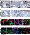
Different pattern of pulmonary metastases formed by MDA/pcDNA and MDA/VEGF-C cells. A, H&E-stained lung sections. MDA/pcDNA metastases localize in the lung parenchyma distant from the small (A1–A2) and large (A3–A4) bronchi. MDA/VEGF-C metastases surround small (A5–A6) and large (A7–A8) bronchi. B, immunofluorescent staining for α-SMA (red) showing MDA/pcDNA metastases in the alveolar region away from bronchi (B1–B2). MDA/VEGF-C metastases are adjacent to the small and large bronchi (B3–B4). b, bronchi; t, tumor (green, GFP). Scale bars, 200 μm (A1, A3, A5, A7), 100 μm (B1–B4), and 50 μm (A2, A4, A6, A8).
Figure 3
Figure 3
Pulmonary tumor emboli are characteristic of MDA/VEGF-C metastases. A, MDA/VEGF-C tumor cells present in pulmonary arteries (arrows) associated with the airways, stained with α-SMA. B, MDA/pcDNA metastases are not found in pulmonary arteries (arrows; B1–B2) nor in veins (v; B3). C, confocal images of MDA/VEGF-C tumor emboli in the pulmonary arteries. C1, metastases labeled with RFP (red). C2, immunofluorescent staining for α-SMA (red) showing tumor embolus (GFP, green) in a small artery (arrow) adjacent to the bronchus (b). C3, staining for blood-endothelial marker CD34 (purple) showing small artery (arrow) with tumor embolus and pulmonary capillaries. C4, large metastatic lesion (GFP, green) involving pulmonary arteries (α-SMA, red, arrows) and the surrounding peribronchial interstitium. D, MDA/pcDNA metastases (GFP, green) are generally distant from the large vasculature (D1). Large pulmonary vessels (arrow) do not contain MDA/pcDNA tumor emboli (D2-D3). Red, a-SMA; purple, CD34. D4–D5, tumor cells (green, arrowheads) localized within pulmonary capillaries (vWF, red). Blue, Hoechst nuclear stain; b, bronchi; t, tumor cells. Data are representative of at least three experiments. Scale bars, 200 μm (A–B), 150 μm (C1), 50 μm (C2–C4, D1–D3), and 25 μm (D4–D5).
Figure 4
Figure 4
Confocal analysis of metastases in pulmonary lymphatics adjacent to the airways, arteries, and veins. A, intralymphatic metastases of MDA/VEGF-C cells adjacent to the airways. Immunofluorescent staining for VEGFR-3 (A1, red), LYVE-1 (A2, red), and podoplanin (A3, red) showing metastases (GFP, green) in lymphatics adjacent to the bronchi (b). A3, α-SMA staining (blue) shows smooth muscle of the bronchial wall. A4-A5, three-dimensional reconstruction of intralymphatic metastases obtained by confocal imaging of 30-μm-thick lung section immunostained with LYVE-1 (red). Transversal (A4) and longitudinal projection (A5). B, metastases in the lymphatics adjacent to the veins (v; B1) and pulmonary arteries (a; B2–B4). Red, podoplanin; blue, α-SMA. Note that pulmonary vein does not contain tumor cells, whereas pulmonary artery and lymphatics are filled with tumor. B4, typical large metastatic lesion in the peribronchial area localizes in dilated lymphatic vessels and in pulmonary artery. C, immunohistochemical staining of lung serial sections for α-SMA and LYVE-1. Collecting lymphatic vessels containing large tumor mass (arrows), adjacent to the bronchus (C1–C2) and the pulmonary vein (C3–C4). Blue or gray, cell nuclei, Hoechst. Scale bar, 100 μm; except in A3, A4, and A5, 50 μm.
Figure 5
Figure 5
Evolution of lymphangiogenesis associated with expansion of metastases. A, immunofluorescent staining for VEGFR-3 (red) showing lymphatic vessels in relation to the MDA/VEGF-C metastases. A1-A3, small metastatic nodule (t, green) next to the lymphatic vessel. Note that during early stages of metastases lymphatics are not changed in number or appearance. A4-A6, dilated lymphatics (arrows) surround larger metastatic lesions and new lymphatics line the edge of metastases. A7-A9, large metastatic lesion infiltrated with lymphatics; many lymphatics contain tumor cells. A10-A12, drastic expansion of the lymphatic network and extreme dilation of lymphatics are associated with very large metastases. Note that metastases and the associated lymphatics always localize in the proximity of the airways. Green, GFP-labeled MDA/VEGF-C metastases; blue, Hoechst nuclear stain; red, VEGFR-3; t, tumor cells; b, bronchi; arrows, dilated lymphatic vessels. Scale bar, 100 μm. B, quantification of the total lymphatic vessel area in the lungs bearing metastases (left). Quantification of the lymphatic vessel area per metastatic foci (right). **, P < 0.01.
Figure 6
Figure 6
VEGF-C expression in metastases of patients with breast adenocarcinoma presenting as lymphangitic carcinomatosis. A, immunohistochemical staining of the lungs for the lymphatic marker D2-40 shows large lymphatics adjacent to the bronchiole and pulmonary artery filled with metastatic breast carcinoma cells (A1–A2). A3, serial section stained for VEGF-C. B, serial sections of another metastatic lesion in the lymphatics adjacent to the pulmonary blood vessel. B1, H&E; B2, lymphatic marker D2-40; B3, VEGF-C. Note massive involvement of lymphatics with the tumor and strong expression of VEGF-C by the metastatic cells. C, metastases within the lung parenchyma from the same patient, stained for VEGF-C (C1–C2). The metastatic tumor deposits appear as moderately differentiated adenocarcinoma with a surrounding desmoplastic reaction. C3, lung metastases of a poorly differentiated adenocarcinoma. Note low levels of VEGF-C expression in the parenchymal metastases and strong expression of VEGF-C by metastases localized in the lymphatic vessels. Arrows, lymphatic vessels; t, tumor cells; b, bronchi. Scale bars, 200 μm (A1, B1–B3, C1–C3) and 100 μm (A2–A3).

References

    1. Tomashefski JF, Dail DH. Dail and Hammar's Pulmonary Pathology. 3rd ed. Springer; New York: 2008.
    1. Alitalo K, Carmeliet P. Molecular mechanisms of lymphangiogenesis in health and disease. Cancer Cell. 2002;1:219–27. - PubMed
    1. Pepper MS, Tille JC, Nisato R, Skobe M. Lymphangiogenesis and tumor metastasis. Cell Tissue Res. 2003;314:167–77. - PubMed
    1. Joukov V, Pajusola K, Kaipainen A, et al. A novel vascular endothelial growth factor, VEGF-C, is a ligand for the Flt4 (VEGFR-3) and KDR (VEGFR-2) receptor tyrosine kinases. EMBO J. 1996;15:290–98. - PMC - PubMed
    1. Lee J, Gray A, Yuan J, Luoh SM, Avraham H, Wood WI. Vascular endothelial growth factor-related protein: a ligand and specific activator of the tyrosine kinase receptor Flt4. Proc Natl Acad Sci U S A. 1996;93:1988–92. - PMC - PubMed

Publication types

MeSH terms

Substances