. 2010 Mar 1;70(5):2064-74.
doi: 10.1158/0008-5472.CAN-09-2684. Epub 2010 Feb 23.

Protein kinase Ciota is required for pancreatic cancer cell transformed growth and tumorigenesis

Affiliations

Protein kinase Ciota is required for pancreatic cancer cell transformed growth and tumorigenesis

Michele L Scotti et al. Cancer Res. .

Abstract

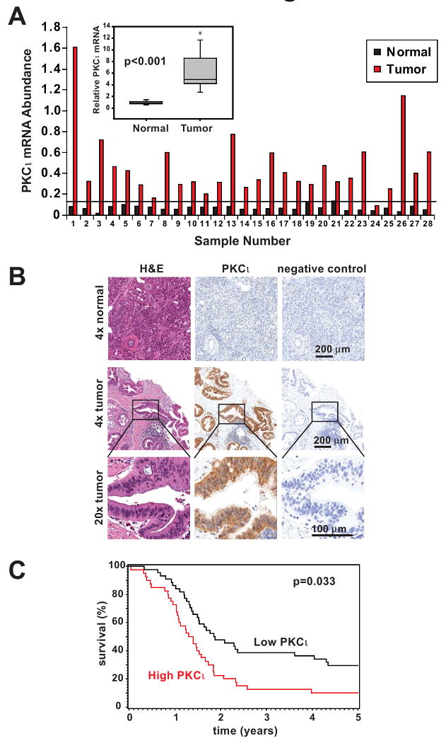
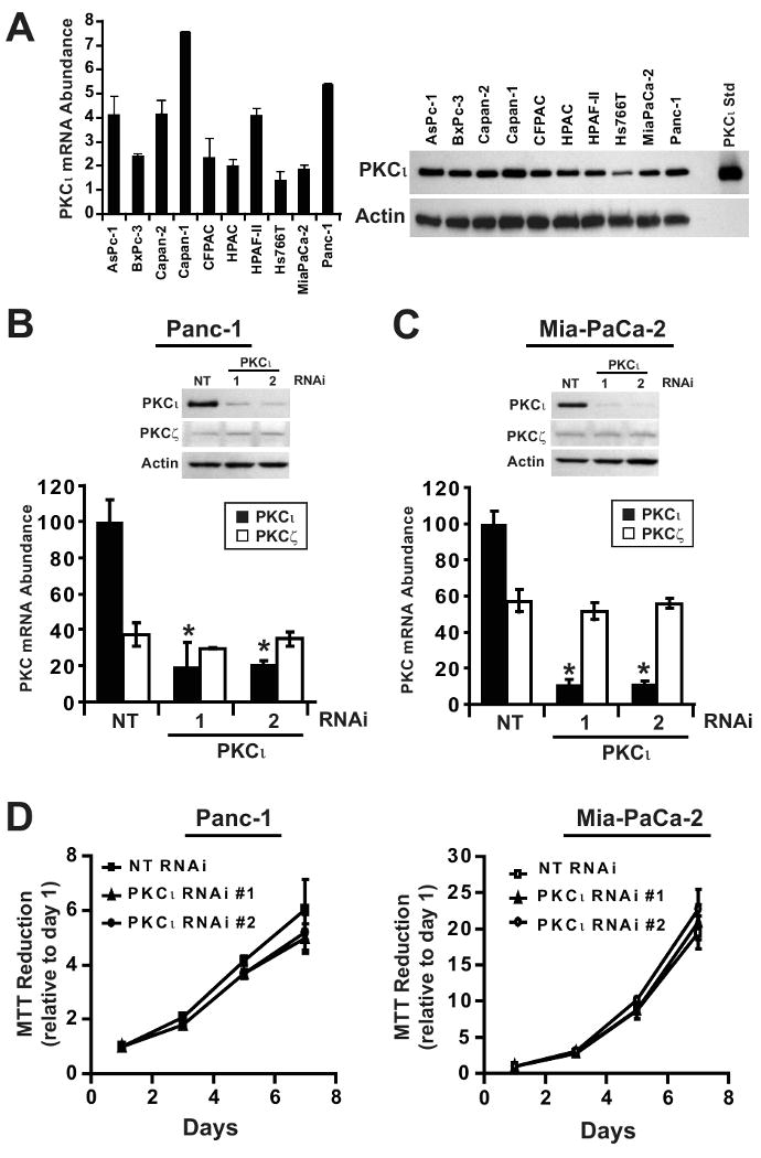
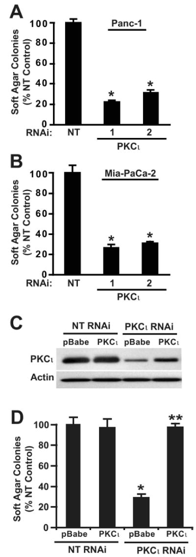
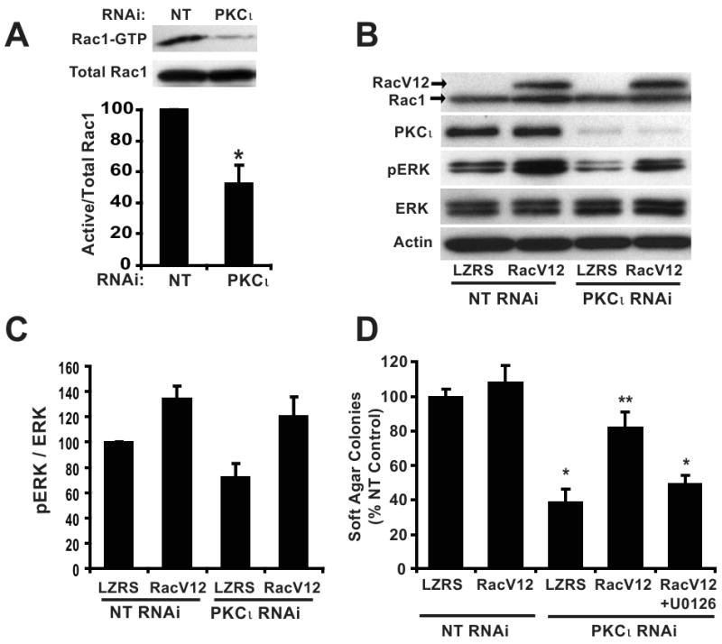
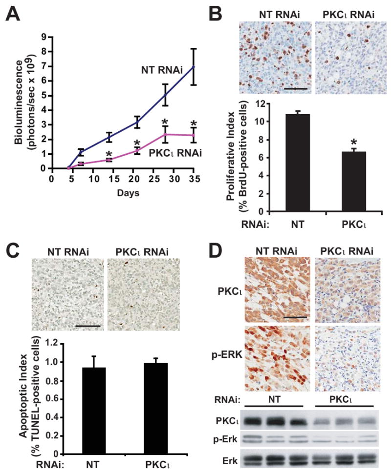
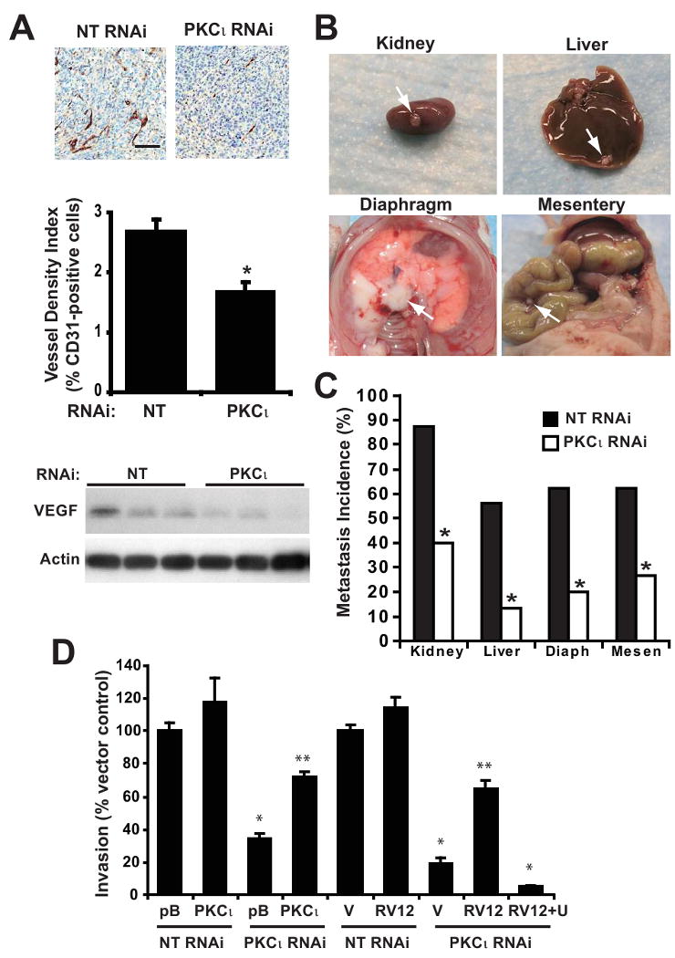
Pancreatic cancer is the fourth leading cause of cancer deaths in the United States, with an overall 5-year survival rate of <5%. Pancreatic ductal adenocarcinoma (PDAC), the most common form of pancreatic cancer, is highly resistant to conventional chemotherapies, underscoring the critical need for new molecular targets for pancreatic cancer chemotherapy. The KRAS proto-oncogene is mutated in >90% of PDAC. Protein kinase Ciota (PKCiota) is required for the oncogenic Ras-mediated transformed growth of lung cancer and intestinal epithelial cells. However, little is known about the role of PKCiota in pancreatic cancer. In this study, we evaluated the expression of PKCiota in human pancreatic cancer and the requirement for PKCiota for the transformed growth and tumorigenicity of PDAC cells. We find that PKCiota is significantly overexpressed in human pancreatic cancer, and high PKCiota expression correlates with poor patient survival. Inhibition of PKCiota expression blocks PDAC cell transformed growth in vitro and tumorigenicity in vivo. Inhibition of PKCiota expression in pancreatic tumors also significantly reduces tumor angiogenesis and metastasis. Analysis of downstream PKCiota effectors implicates the Rac1-MEK/ERK1/2 signaling axis in PKCiota-mediated transformed growth and cellular invasion. Taken together, our data show a required role for PKCiota in the transformed growth of pancreatic cancer cells and reveal a novel role for PKCiota in pancreatic cancer cell metastasis and angiogenesis in vivo. Our results strongly indicate that PKCiota will be an effective target for pancreatic cancer therapy.


Figures

Figure 1
Figure 1. PKCι is highly expressed in human pancreatic cancer and correlates with poor survival in PDAC patients
A) qPCR analysis of PKCι mRNA expression in 28 matched human pancreatic tumor and adjacent non-tumor pancreas. Data were normalized to 18S RNA abundance (× 104) to control for RNA concentration. Horizontal line indicates 2 standard deviations above the mean PKCι mRNA abundance in adjacent non-tumor pancreas samples. Inset: PKCι mRNA expression is significantly increased in tumors compared to matched non-tumor pancreas tissue. Average fold increase in PKCι mRNA abundance in tumor/matched non-tumor is plotted. B) Representative images of IHC detection of PKCι expression in formalin-fixed human pancreatic adenocarcinoma and normal pancreas. H&E staining and negative control secondary antibody staining are also shown in serial sections. C) Kaplan-Meier survival curves. PDAC patient tumors were analyzed by IHC for PKCι expression and divided into high (red line) and low (black line) expression groups as described in Materials and Methods.
Figure 2
Figure 2. PKCι is highly expressed in PDAC cell lines and is not required for anchorage-dependent (non-transformed) growth of PDAC cells
A) Left, qPCR analysis of PKCι mRNA expression in ten human pancreatic cancer cell lines. mRNA abundance is normalized to GAPDH (× 102), n=3. Right, Immunoblot analysis of ten human pancreatic cancer cell lines for expression of PKCι and β-actin. qPCR analysis of PKCι and PKCζ mRNA expression in B) Panc-1 and C) MiaPaCa-2 stably carrying either non target (NT), PKCι-specific RNAi constructs (PKCι #1) or (PKCι #2). PKC mRNA abundance is normalized to GAPDH and presented relative to PKCι in NT RNAi cells. Insets, Immunoblot analysis of PKCι, PKCζ and β-actin protein expression in B) Panc-1 and C) MiaPaCa-2 NT or PKCι-RNAi (PKCι#1 and PKCι#2) constructs. D) Anchorage-dependent growth in Panc-1 (left) and MiaPaCa-2 (right) stably carrying either NT or PKCι-RNAi (PKCι#1 and PKCι#2) was determined by MTT colorimetric assay. Analysis was performed in triplicate and represents two independent experiments.
Figure 3
Figure 3. PKCι is required for anchorage-independent growth of PDAC cells
Soft agar colony formation of A) Panc-1 and B) MiaPaCa-2 cells with NT or PKCι-RNAi (PKCι#1 and PKCι#2) constructs. *= significantly different than NT. C) Immunoblot analysis of PKCι expression in Panc-1 cells co-transfected with RNAi (NT or PKCι) and control vector (pBabe) or vector expressing wild type PKCι (PKCι). D) Re-expression of PKCι overcomes the inhibitory effect of PKCι RNAi on soft agar colony formation. *= significantly different than control (NT & pBabe), **= significantly different than PKCι RNAi & pBabe. Mean +/-SEM is plotted and represents two independent experiments.
Figure 4
Figure 4. Constitutively active Rac1 (RacV12) recovers transformed growth of PKCι RNAi PDAC cells in a MEK-dependent manner
A) Panc-1 cells stably expressing NT or PKCι RNAi were assayed for Rac1 activity. Top panel, (Active) Rac1-GTP was precipitated from cell extracts with PAK-1 PBD agarose. Immunoblot analysis of precipitates and total cellular extracts (total Rac1) was performed using an anti-Rac1 antibody. Bottom panel, Quantitative, densitometric analysis of relative Rac1 activity (active Rac1/total Rac1). Mean +/- SEM is plotted, n=3. B) Panc-1 cells co-transfected with RNAi (NT or PKCι) and control vector (LZRS) or vector expressing RacV12 were subject to immunoblot analysis for expression of Rac1, PKCι, p-ERK1/2 (Thr202/Tyr204), ERK1/2 and actin as a loading control. Arrows indicate migration of endogenous Rac1 and slower migrating myc-tagged RacV12. C) Quantitation of densitometric analysis of relative p-ERK1/2 to ERK1/2 expression. Mean +/- SEM is plotted, n=3. D) Expression of RacV12 recovers the inhibitory effect of PKCι RNAi on soft agar colony formation and requires MEK activity. +U0126= 10 μM U0126 was included in the assay media and agar; *= significantly different than control (NT & LZRS) and RacV12 reconstituted (PKCι RNAi & RacV12); **= significantly different than PKCι KD (PKCι RNAi & LZRS) and MEK-inhibited (PKCι RNAi & RacV12+U0126). Mean +/-SEM is plotted and represents two independent experiments.
Figure 5
Figure 5. Inhibition of PKCι blocks orthotopic pancreatic tumor proliferation and proliferative signaling
A) Tumor growth was monitored by bioluminescence (total flux, photons/sec) detected by IVIS imaging of orthotopic Panc-1 NT versus Panc-1 PKCι RNAi pancreatic tumors in live, anesthetized mice at weekly intervals after tumor implantation. n=15, 16/group. *= significantly different than NT RNAi tumors. B) Top: Immunohistochemical analysis of BrdUrd incorporation. Bar=100μm. Bottom: Quantitative analysis of BrdUrd incorporation into Panc-1 tumors. Mean+/-SEM is plotted. C) Top: Immunohistochemical detection of TUNEL staining in representative tumors. Bar=100μm. Bottom: Quantitative analysis of TUNEL staining. Mean+/-SEM is plotted. D) Representative images of IHC detection of PKCι and p-ERK1/2 (Thr 202/Tyr 204) in NT and PKCι RNAi tumors. Bar=100μm. Representative immunoblot analysis of PKCι, p-ERK1/2 (Thr 202/Tyr 204) and ERK1/2 in Panc-1 NT and PKCι RNAi orthotopic pancreatic tumors. Equivalent amounts of protein from each tumor sample were analyzed.
Figure 6
Figure 6. Inhibition of PKCι blocks PDAC angiogenesis and metastasis
A) Top: Immunohistochemical detection of CD31 staining. Bar=100μm. Middle: Quantitative analysis of CD31 positive staining in Panc-1 tumors, calculated as the ratio of CD31-positive pixels to the sum of all pixels. Mean+/-SEM is plotted. Bottom: Representative immunoblot analysis of VEGF and actin in Panc-1 NT and PKCι RNAi orthotopic pancreatic tumors. B) Representative images of tumor metastases to various organs. C) Percent of orthotopic Panc-1 NT and PKCι RNAi pancreatic tumors that metastasized to various organs is plotted. *= significantly different than NT RNAi tumors. D) Panc-1 cells were assayed for cellular invasion through Matrigel-coated chambers as described in Materials and Methods. pB=pBABE control vector; V=LZRS control vector; RV12= RacV12; RV12+U= RacV12 treated with 10 μM U1026; *= significantly different than control (NT & pB) or (NT & V), **= significantly different than (PKCι RNAi & pB) or (PKCι RNAi & V) or (PKCι RNAi & RV12 +U). Mean +/-SEM is plotted and represents two independent experiments.

References

    1. Lebedeva IV, Sarkar D, Su ZZ, et al. Molecular target-based therapy of pancreatic cancer. Cancer Res. 2006;66:2403–13. - PubMed
    1. Klimstra DS, Longnecker DS. K-ras mutations in pancreatic ductal proliferative lesions. Am J Pathol. 1994;145:1547–50. - PMC - PubMed
    1. Aoki K, Yoshida T, Matsumoto N, Ide H, Sugimura T, Terada M. Suppression of Ki-ras p21 levels leading to growth inhibition of pancreatic cancer cell lines with Ki-ras mutation but not those without Ki-ras mutation. Mol Carcinog. 1997;20:251–8. - PubMed
    1. Saad ED, Hoff PM. Molecular-targeted agents in pancreatic cancer. Cancer Control. 2004;11:32–8. - PubMed
    1. Murray NR, Jamieson L, Yu W, et al. Protein kinase C{iota} is required for Ras transformation and colon carcinogenesis in vivo. J Cell Biol. 2004;164:797–802. - PMC - PubMed

Publication types

MeSH terms