. 2010 Feb 23;3(110):ra14.
doi: 10.1126/scisignal.2000467.

DNA-PKcs controls an endosomal signaling pathway for a proinflammatory response by natural killer cells

Affiliations

DNA-PKcs controls an endosomal signaling pathway for a proinflammatory response by natural killer cells

Sumati Rajagopalan et al. Sci Signal. .

Abstract

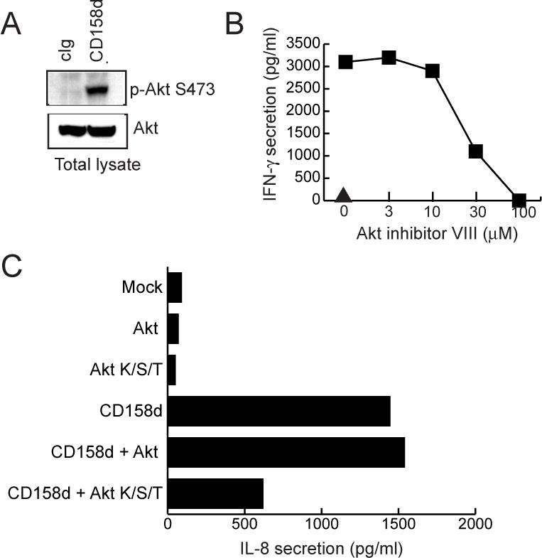
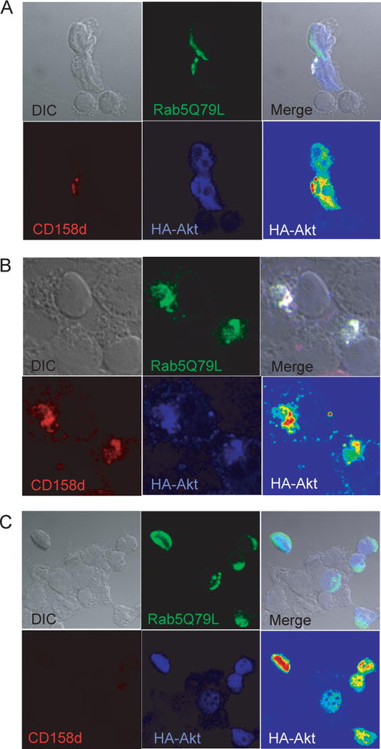
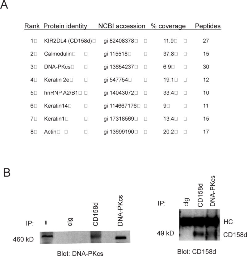
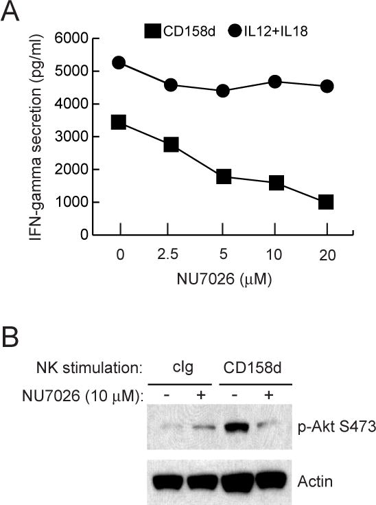
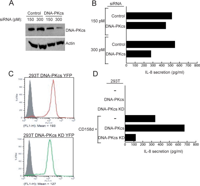
Endosomes are emerging as specialized signaling compartments that endow receptors with distinct signaling properties. The diversity of endosomal signaling pathways and their contribution to various biological responses is still unclear. CD158d, which is also known as the killer cell immunoglobulin-like receptor (KIR) 2DL4 (KIR2DL4), is an endosome-resident receptor in natural killer (NK) cells that stimulates the release of a unique set of proinflammatory and proangiogenic mediators in response to soluble human leukocyte antigen G (HLA-G). Here, we identified the CD158d signaling cascade. In response to soluble agonist antibody or soluble HLA-G, signaling by CD158d was dependent on the activation of nuclear factor kappaB (NF-kappaB) and the serine-threonine kinase Akt. CD158d associated with the catalytic subunit of DNA-dependent protein kinase (DNA-PKcs), promoted the recruitment of Akt to endosomes, and stimulated the DNA-PKcs-dependent phosphorylation of Akt. The sequential requirement for DNA-PKcs, Akt, and NF-kappaB in signaling by CD158d delineates a previously uncharacterized endosomal signaling pathway for a proinflammatory response in NK cells.


Figures

Fig. 1
Fig. 1
Determinants of endosomal localization and signaling by CD158d. (A) Schematic representation of CD158d, gp49B, and chimeric variants. Each construct carries an HA epitope tag at the N-terminus. EC: extracellular domain; TM: transmembrane region; CYT: cytoplasmic tail. (B) Receptor localization by confocal microscopy analysis. 293T cells transfected with the indicated constructs were stained with an anti-HA tag Ab after 48 h. (C) IL-8 secretion induced by the indicated plasmids in transfected 293T cells.
Fig. 2
Fig. 2
NF-κB activation is involved in signaling by CD158d. (A) Activity of the indicated reporter plasmids in 293T cells transfected with control vector (grey bars) or with a plasmid encoding CD158d (black bars). Stimulation with TNF-α (10 ng/ml) during the last 5 h of culture was used as a positive control for NF-κB activation. Results are presented as the ratio of Firefly luciferase activity to Renilla luciferase activity (F/R). The data are representative of three independent experiments. (B) NF-κB reporter activity in cells transfected with the indicated plasmids, as in (A). TNF-α (10 ng/ml) was present where indicated during the last 5 h of culture. The data are representative of three independent experiments. (C) IL-8 secretion by 293T cells transfected with plasmid encoding CD158d together with increasing amounts of a plasmid encoding the constitutive inhibitor CI-IκBα. Supernatants were tested for IL-8 48 h after transfection. The data are representative of three experiments. (D) Upper panels: IκBα phosphorylation in resting NK cells stimulated with 10 μg/ml of control antibody (cIg), CD158d mAb, or sHLA-G for 16 h. Cell lysates were immunoblotted for phospho-IκBα and tubulin (loading control). Lower panels: Translocation of p65 into the nucleus of resting NK cells stimulated with 10 μg/ml of cIg, CD158d mAb, or sHLA-G for 16 h. Cytoplasmic (cyt) and nuclear (nuc) fractions were lysed and analyzed by immunoblotting for p65, tubulin (a cytoplasmic marker), and histone deacetylase (HDAC, a nuclear marker). The data are representative of experiments done in resting NK cells from three different donors.
Fig. 3
Fig. 3
CD158d signaling induces Akt phosphorylation. (A) Inhibition of IFN-γ release by resting NK cells with PP2 or wortmannin at the indicated concentrations. Inhibitors were added for 1 h at 37°C before activation with CD158d mAb or with a combination of IL-12 (10 ng/ml) and IL-18 (25 ng/ml) for 16 h in the continuing presence of the inhibitor. The triangle represents the IFN-γ secretion induced by isotype-matched control antibody. (B) Kinase phosphorylation profile during CD158d signaling. Lysates of 293T cells transfected with either CD158d or tail-deleted receptor (CD158d-TR) were assayed with the Human Phospho-Kinase Profiler Array (RnD Systems #ARY002). (C) Signals from scanned X-ray film images in (B) were plotted as a function of average pixel density. Numbers represent corresponding data on the scan and the graph.
Fig. 4
Fig. 4
Akt activation is required for CD158d signaling. (A) Akt phosphorylation in resting NK cells stimulated with 10 μg/ml of control antibody (cIg), CD158d mAb, and sHLA-G for 16 h. Cell lysates were immunoblotted for Akt S473 phosphorylation and for tubulin (loading control). (B) IFN-γ and IL-8 secretion by resting NK cells treated with Akt inhibitor VIII at 37°C for 1 h before stimulation and for 16 h during stimulation with IgG CD158d mAb. The triangle denotes IFN-γ and IL-8 secretion in the presence of isotype-matched control mAb. (C) Inhibition of Akt phosphorylation by resting NK cells with Bafilomycin A1 (1 μM) or Dynasore (50 μM). Inhibitors were added for 1 h at 37°C before activation with CD158d mAb for 16 h in the continued presence of inhibitor. Cells lysates were immunoblotted for Akt phosphorylation at S473 and for tubulin (loading control). Data shown in A-C are representative of experiments done using resting NK cells from three different donors. (D) Dominant-negative Akt (Akt K/S/T) impairs CD158d function. IL-8 secretion by 293T cells transfected with the indicated plasmids. The average inhibition of IFN-γ secretion by Akt K/S/T, relative to wild type Akt, in 4 independent experiments was 62.15% ± 3.65.
Fig. 5
Fig. 5
Akt is recruited to endosomes containing CD158d. (A) Rab5Q79L enhances CD158d function. IL-8 secretion by 293T cells transfected with the indicated plasmids for 48 h. IL-8 secretion collected during the last 16 h was determined. (B) A single confocal section of CD158d-negative 293T cells co-transfected with Rab5Q79L-GFP (green) and HA-Akt (blue). In 21 CD158d-negative cells analyzed, no co-localization of Akt with Rab5Q79L-positive endosomes was observed. The average colocalization coefficient for Akt and Rab5Q79L in the transfected cells in the field shown was 0.128 ± 0.142. (C, D) A single confocal section of 293T cells co-transfected with Rab5Q79L-GFP (green), CD158d (red), and HA-Akt (blue). An intensity scale (red=high, blue=low) is also displayed for HA-Akt in the bottom right panel. Cells were loaded with Cy3-labeled CD158d mAb for 2 h. Akt was labeled post-fixation and permeabilization with anti-HA mAb followed by Alexa-647 secondary Ab. For comparison, note that in the CD158d-negative cell at the top in (C), Akt did not co-localize with Rab5Q79L-positive endosomes. Additional images are shown in Figure S4. The colocalization coefficient for Akt and Rab5Q79L in the cells in Fig. 5C is as follows: CD158d-negative cell, top: 0.115; CD158d-positive cell, bottom: 0.799. Fig. 5D: 0.704 and 0.711 for the cells on the left and right, respectively. See Fig. S4 and S5 for additional colocalization image displays and profiles.
Fig. 6
Fig. 6
DNA-PKcs is associated with CD158d. (A) Proteins associated with TAP-tagged CD158d identified by μLC/MS/MS. Rank is based on the intensity of the MS/MS spectra. The number of unique peptides in the MS/MS spectra is shown. (B) Association of DNA-PKcs with CD158d in NK cells. NKL cells were stimulated with IgM CD158d mAbs for 2 h. Lysates without immunoprecipitation (−) or immunoprecipitated with control Ab (cIg) or Abs to CD158d and DNA-PKcs, as indicated, were immunoblotted for DNA-PKcs (left panel) and CD158d (right panel).
Fig. 7
Fig. 7
Inhibition of DNA-PKcs blocks CD158d signaling. (A) IFN-γ secretion by resting NK cells stimulated with IgG CD158b mAb or with IL-12 (10 ng/ml) and IL-18 (25 ng/ml) for 16 h. IL-8 secretion by resting NK cells stimulated with CD158d mAb is also shown. NK cells were treated with the indicated concentration of NU7026 for 1 h at 37°C before stimulation and during the 16 h of stimulation. The triangle denotes secretion in the presence of isotype-matched control antibody. (B) Akt S473 phosphorylation detected by immunoblotting in lysates of resting NK cells stimulated with cIg or CD158d mAb for 16 h. Where indicated (+), cells were pre-incubated with 10 μM NU7026 for 1 h prior to stimulation and during the 16 h of stimulation. Blots were re-probed for actin.
Fig. 8
Fig. 8
DNA-PKcs activity is required for CD158d signaling. (A) siRNA knockdown of DNA-PKcs in 293T-CD158d-GFP cells. 48 h after transfection with siRNA cell lysates were immunoblotted for DNA-PKcs and re-probed for actin. (B) IL-8 secretion by 293T-CD158d-GFP cells 48 h after transfection with the indicated siRNA. The average inhibition of IL-8 secretion by DNA-PKcs siRNA at 300 pM in 3 independent experiments was 49.47% ± 8.06%. (C) Stable expression of DNA-PKcs-YFP and a kinase-dead (KD) mutant of DNA-PKcs-YFP in 293T cells. The grey profile represents untransfected 293T cells. (D) IL-8 secretion by 293T cells, 293T-DNA-PKcs-YFP and 293T-DNA-PKcs-KD-YFP cells. As indicated, these cells were either not transfected or transiently transfected with a CD158d plasmid for 48 h. All cells were transfected with a Renilla luciferase reporter construct to normalize for transfection efficiency in the different cell lines. The data is representative of three independent experiments.

References

    1. Miaczynska M, Pelkmans L, Zerial M. Not just a sink: endosomes in control of signal transduction. Curr Opin Cell Biol. 2004;16:400–406. - PubMed
    1. Polo S, Di Fiore PP. Endocytosis conducts the cell signaling orchestra. Cell. 2006;124:897–900. - PubMed
    1. von Zastrow M, Sorkin A. Signaling on the endocytic pathway. Curr Opin Cell Biol. 2007;19:436–445. - PMC - PubMed
    1. Hoeller D, Volarevic S, Dikic I. Compartmentalization of growth factor receptor signalling. Curr Opin Cell Biol. 2005;17:107–111. - PubMed
    1. Sorkin A, Von Zastrow M. Signal transduction and endocytosis: close encounters of many kinds. Nat Rev Mol Cell Biol. 2002;3:600–614. - PubMed

Publication types

MeSH terms

LinkOut - more resources