

American ginseng suppresses colitis through p53-mediated apoptosis of inflammatory cells
- PMID: 20179294
- PMCID: PMC2833220
- DOI: 10.1158/1940-6207.CAPR-09-0116
American ginseng suppresses colitis through p53-mediated apoptosis of inflammatory cells
Abstract
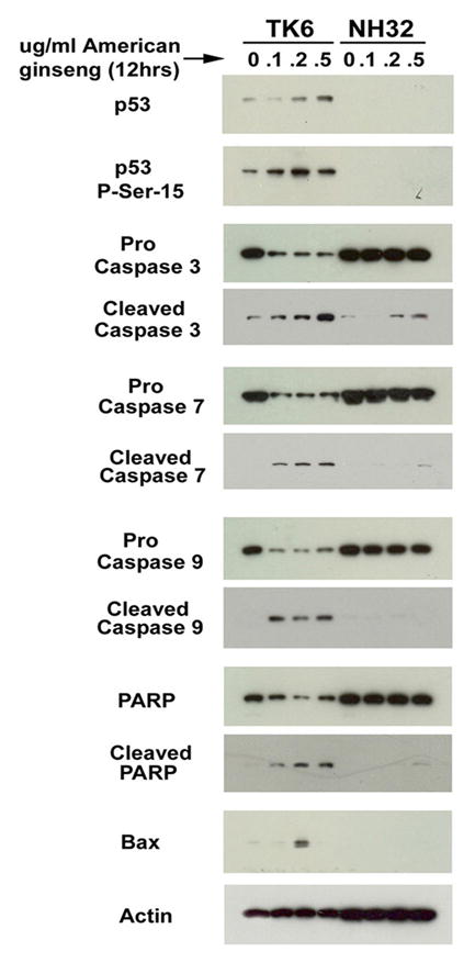
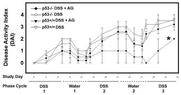
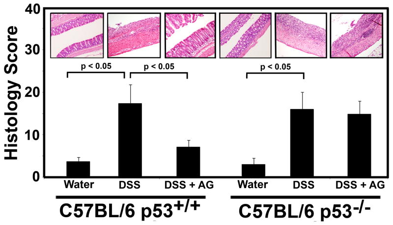
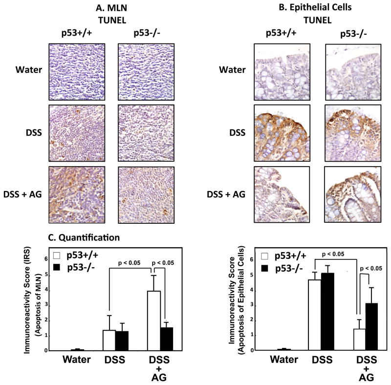
Ulcerative colitis is a dynamic, chronic inflammatory condition associated with an increased colon cancer risk. Inflammatory cell apoptosis is a key mechanism regulating ulcerative colitis. American ginseng (AG) is a putative antioxidant that can suppress hyperactive immune cells. We have recently shown that AG can prevent and treat mouse colitis. Because p53 levels are elevated in inflammatory cells in both mouse and human colitis, we tested the hypothesis that AG protects from colitis by driving inflammatory cell apoptosis through a p53 mechanism. We used isogenic p53(+/+) and p53(-/-) inflammatory cell lines as well as primary CD4(+)/CD25(-) effector T cells from p53(+/+) and p53(-/-) mice to show that AG drives apoptosis in a p53-dependent manner. Moreover, we used a dextran sulfate sodium (DSS) model of colitis in C57BL/6 p53(+/+) and p53(-/-) mice to test whether the protective effect of AG against colitis is p53 dependent. Data indicate that AG induces apoptosis in p53(+/+) but not in isogenic p53(-/-) cells in vitro. In vivo, C57BL/6 p53(+/+) mice are responsive to the protective effects of AG against DSS-induced colitis, whereas AG fails to protect from colitis in p53(-/-) mice. Furthermore, terminal deoxynucleotidyl transferase-mediated dUTP nick end labeling of inflammatory cells within the colonic mesenteric lymph nodes is elevated in p53(+/+) mice consuming DSS + AG but not in p53(-/-) mice consuming DSS + AG. Results are consistent with our in vitro data and with the hypothesis that AG drives inflammatory cell apoptosis in vivo, providing a mechanism by which AG protects from colitis in this DSS mouse model.
Figures
Similar articles
-
A hexane fraction of American ginseng suppresses mouse colitis and associated colon cancer: anti-inflammatory and proapoptotic mechanisms.Cancer Prev Res (Phila). 2012 Apr;5(4):685-96. doi: 10.1158/1940-6207.CAPR-11-0421. Epub 2012 Jan 31. Cancer Prev Res (Phila). 2012. PMID: 22293630 Free PMC article.
-
A limited role of p53 on the ability of a Hexane fraction of American ginseng to suppress mouse colitis.J Biomed Biotechnol. 2012;2012:785739. doi: 10.1155/2012/785739. Epub 2012 Jul 30. J Biomed Biotechnol. 2012. PMID: 22899889 Free PMC article.
-
American ginseng suppresses inflammation and DNA damage associated with mouse colitis.Carcinogenesis. 2008 Dec;29(12):2351-9. doi: 10.1093/carcin/bgn211. Epub 2008 Sep 18. Carcinogenesis. 2008. PMID: 18802031 Free PMC article.
-
Mechanistic insight into the ability of American ginseng to suppress colon cancer associated with colitis.Carcinogenesis. 2010 Oct;31(10):1734-41. doi: 10.1093/carcin/bgq163. Epub 2010 Aug 20. Carcinogenesis. 2010. PMID: 20729391 Free PMC article.
-
Ginkgo biloba extract EGb 761 has anti-inflammatory properties and ameliorates colitis in mice by driving effector T cell apoptosis.Carcinogenesis. 2008 Sep;29(9):1799-806. doi: 10.1093/carcin/bgn143. Epub 2008 Jun 20. Carcinogenesis. 2008. PMID: 18567620 Free PMC article.
Cited by
-
American ginseng significantly reduced the progression of high-fat-diet-enhanced colon carcinogenesis in Apc (Min/+) mice.J Ginseng Res. 2015 Jul;39(3):230-7. doi: 10.1016/j.jgr.2014.12.004. Epub 2015 Jan 10. J Ginseng Res. 2015. PMID: 26199554 Free PMC article.
-
Lovastatin Combination Therapy Increases the Survival and Proliferation of Rat Bone Marrow-Derived Mesenchymal Stem Cells Against the Inflammatory Activity of Lipopolysaccharide.Cell Biochem Biophys. 2024 Sep;82(3):2585-2595. doi: 10.1007/s12013-024-01372-z. Epub 2024 Jul 4. Cell Biochem Biophys. 2024. PMID: 38963603
-
Salvia miltiorrhiza (dan shen) significantly ameliorates colon inflammation in dextran sulfate sodium induced colitis.Am J Chin Med. 2013;41(5):1097-108. doi: 10.1142/S0192415X13500742. Am J Chin Med. 2013. PMID: 24117071 Free PMC article.
-
Pharmacological effects of ginseng and ginsenosides on intestinal inflammation and the immune system.Front Immunol. 2024 Apr 18;15:1353614. doi: 10.3389/fimmu.2024.1353614. eCollection 2024. Front Immunol. 2024. PMID: 38698858 Free PMC article. Review.
-
Panaxynol, a bioactive component of American ginseng, targets macrophages and suppresses colitis in mice.Oncotarget. 2020 Jun 2;11(22):2026-2036. doi: 10.18632/oncotarget.27592. eCollection 2020 Jun 2. Oncotarget. 2020. PMID: 32547701 Free PMC article.
References
-
- Sartor RB. Mechanisms of disease: pathogenesis of Crohn’s disease and ulcerative colitis. Nat Clin Pract Gastroenterol Hepatol. 2006;3:390–407. - PubMed
-
- Neuman MG. Immune dysfunction in inflammatory bowel disease. Transl Res. 2007;149:173–186. - PubMed
-
- Hofseth LJ, Hussain SP, Harris CC. p53: 25 years after its discovery. Trends Pharmacol Sci. 2004;25:177–181. - PubMed
-
- Lashner BA, Bauer WM, Rybicki LA, Goldblum JR. Abnormal p53 immunohistochemistry is associated with an increased colorectal cancer-related mortality in patients with ulcerative colitis. Am J Gastroenterol. 2003;98:1423–1427. - PubMed
-
- Alkim C, Savas B, Ensari A, et al. Expression of p53, VEGF, Microvessel Density, and Cyclin-D1 in Noncancerous Tissue of Inflammatory Bowel Disease. Dig Dis Sci. 2008;26:26. - PubMed
Publication types
MeSH terms
Substances
Grants and funding
LinkOut - more resources
Full Text Sources
Medical
Research Materials
Miscellaneous
