. 2010 Mar;3(3):348-58.
doi: 10.1158/1940-6207.CAPR-09-0146. Epub 2010 Feb 23.

Chronic cyclooxygenase-2 inhibition promotes myofibroblast-associated intestinal fibrosis

Affiliations

Chronic cyclooxygenase-2 inhibition promotes myofibroblast-associated intestinal fibrosis

Jennifer S Davids et al. Cancer Prev Res (Phila). 2010 Mar.

Abstract

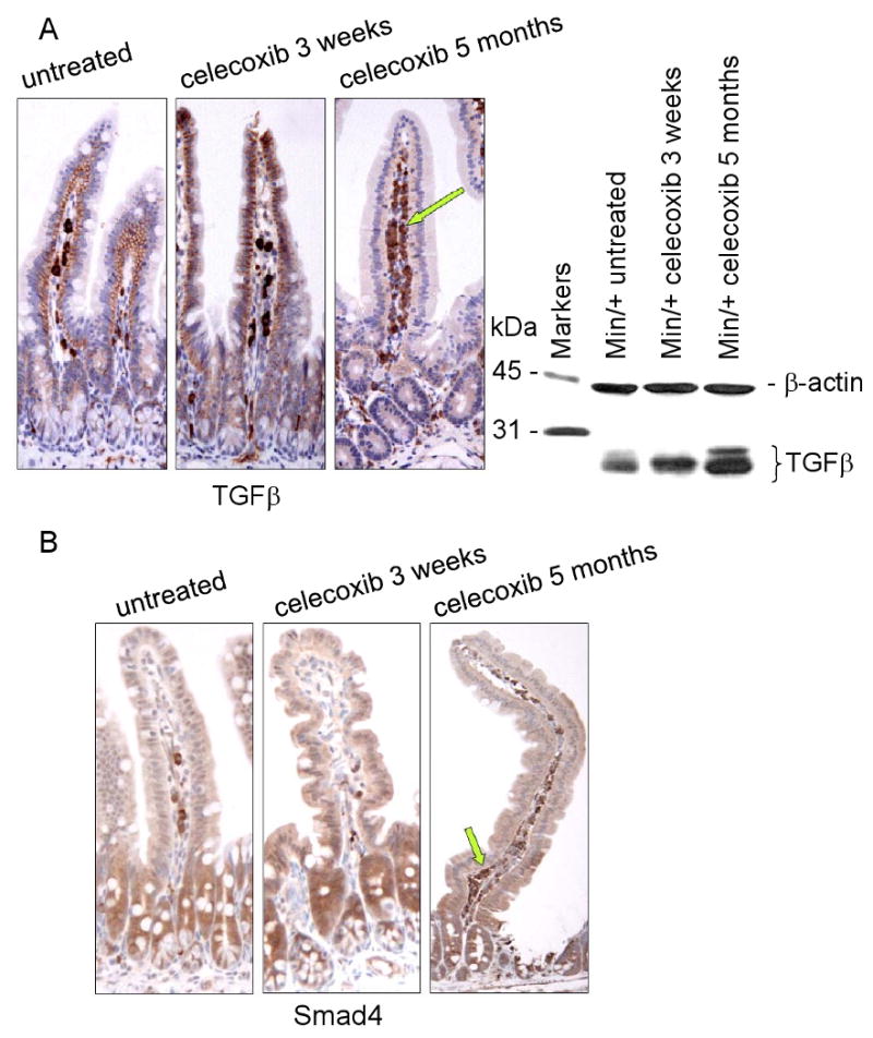
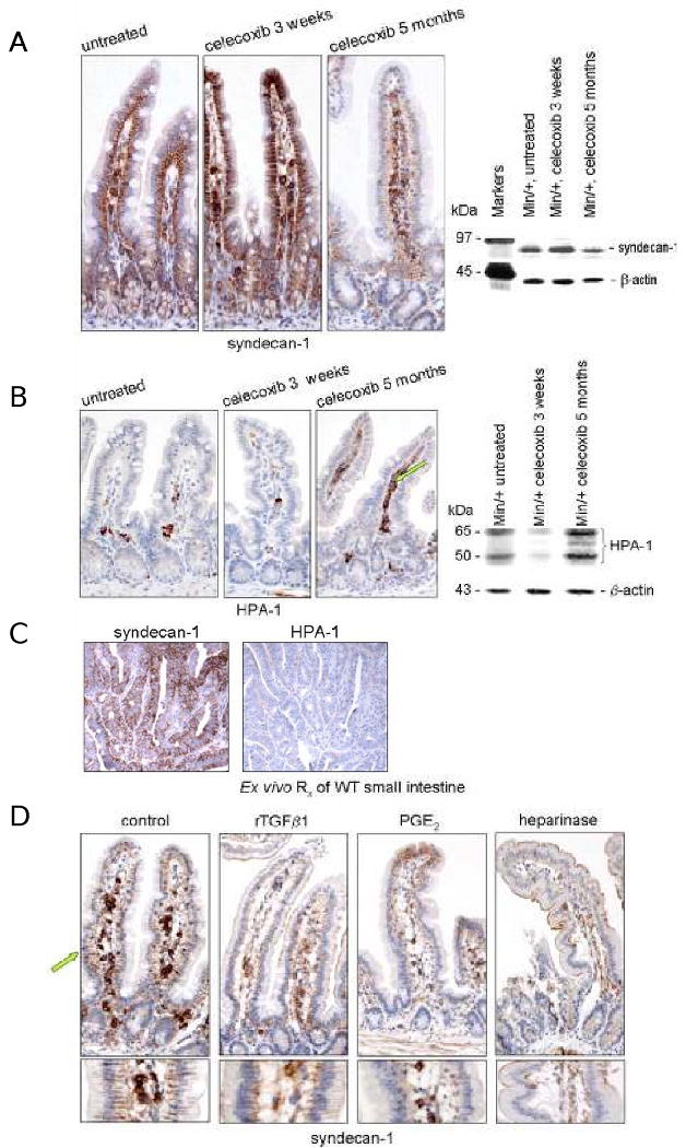
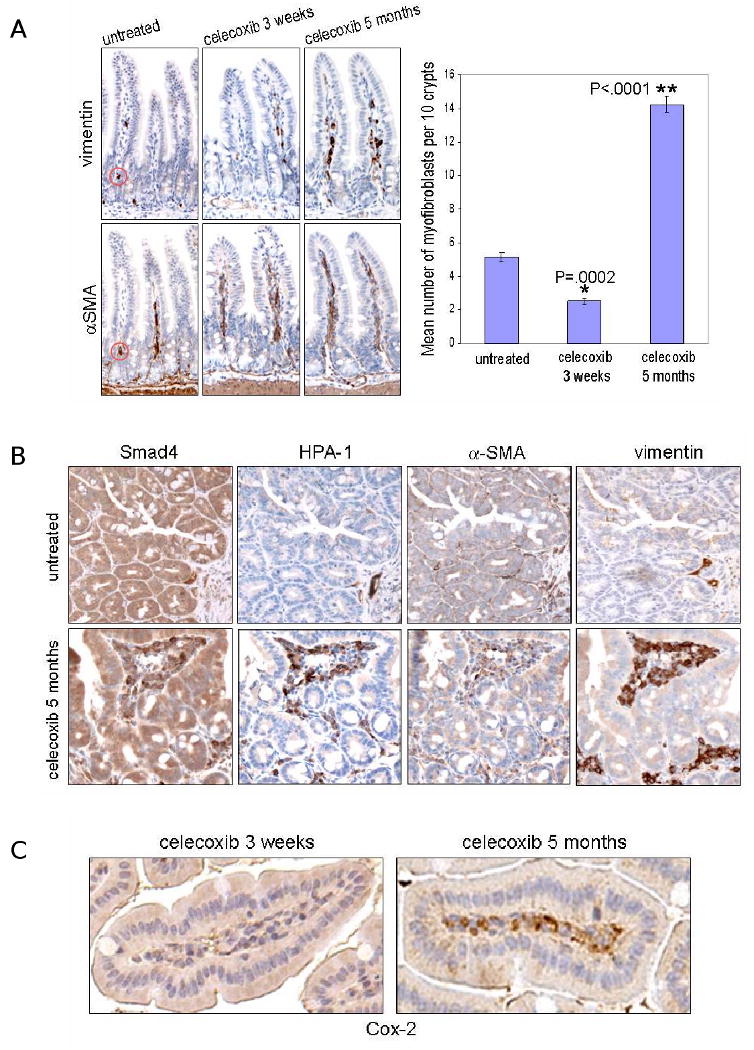
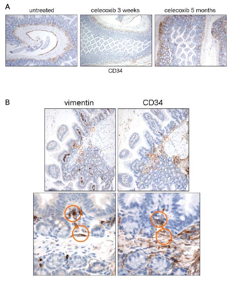
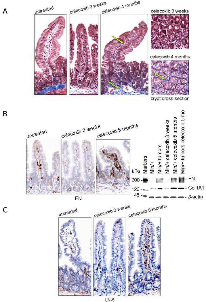
Anti-inflammatory drugs prevent intestinal tumor formation, an activity related to their ability to inhibit inflammatory pathway signaling in the target tissue. We previously showed that treatment of Min/(+) mice with the selective cyclooxygenase-2 (COX-2) inhibitor celecoxib induced rapid tumor regression; however, drug-resistant tumors appeared with long-term treatment. In this study, we investigated whole-tissue changes in inflammatory signaling by studying constituents of the tissue stroma and extracellular matrix. We found that celecoxib resistance was associated with changes in factors regulating autocrine transforming growth factor-beta (TGFbeta) signaling. Chronic drug treatment expanded the population of bone marrow-derived CD34(+) vimentin(+) alphaSMA(-) myofibroblast precursors and alphaSMA(+) vimentin(+) F4/80(-) myofibroblasts in the lamina propria and submucosa, providing a source of increased TGFbeta and COX-2 expression. Membrane constituents regulating TGFbeta availability, including syndecan-1 and heparanase-1, were also modified by chronic treatment in a manner promoting increased TGFbeta signaling. Finally, long-term celecoxib treatment induced tissue fibrosis, as indicated by increased expression of collagen, fibronectin, and laminin in the basement membrane. We conclude that chronic COX-2 inhibition alters TGFbeta signaling in the intestinal mucosa, producing conditions consistent with chronic inflammation.


Figures

Figure 1
Figure 1. Long term celecoxib treatment increases TGFβ signaling in the intestinal stroma
(A) Representative photomicrographs of IHC for TGFβ using sectioned ileum from untreated Min/+ mice, or those treated with celecoxib short (3 weeks) or long term (5 months). Arrow points to positively stained stromal cells in the lamina propria. IB analysis of total cell lysates from control and treated mice measured the expression of TGFβ using 1D11 antibody. The loading control shown below was probed for β-actin using AC-40 antibody. (B) IHC for Smad-4 correlated the presence or absence of membrane-localized TGFβ on enterocytes with low or high TGFβ downstream signaling in the stroma, respectively. Arrows highlight the relative abundance of stained stromal cells. The antibody dilutions used for all IHC performed in this study are provided in Supplementary Table 1. The original magnification on all IHC images was 20×, unless otherwise indicated. All IHC was performed on the complete treatment set in parallel and all experiments used tissues from at least 3 different mice of the same treatment group.
Figure 2
Figure 2. Regulation of TGFβ availability by altered membrane expression of syndecan-1
(A) Representative photomicrographs of IHC for syndecan-1 using sectioned ileum from untreated and treated Min/+ mice. IB analysis of total cell lysates from control and treated mice assessed the steady-state expression of syndecan-1 using 291-2 antibody. (B) Representative photomicrographs of IHC for HPA-1 of sectioned ileum from untreated and treated Min/+ mice. IB analysis for HPA-1 expression used HP3/17 antibody, a probe that detects two isoforms of HPA-1 in intestinal lysates (20). (C) Serial sections of an adenoma from an untreated Min/+ mouse immunostained for syndecan-1 and HPA-1. (D) Representative photomicrographs of sections of ex vivo-treated wild type ileum immunostained for syndecan-1. Tissues were incubated in medium with or without a drug treatment (PGE2, rTGFβ1, or heparinase) prior to formalin-fixing and paraffin-embedding. Treatment conditions were those specified in the Materials and Methods (42).
Figure 3
Figure 3. Chronic celecoxib treatment increased the number of myofibroblasts in Min/+ intestine
(A) Stromal myofibroblasts identified in representative photomicrographs of IHC for vimentin and αSMA of serially sectioned ileum from untreated and treated Min/+ mice. Doubly-positive cells were identified as indicated (red circles). The graph depicts the mean number of myofibroblasts per 10 crypts for the different celecoxib treatment times. N = 260 crypts from Min/+ ileum per treatment group; error bars represent SEM. (B) Adenomas from long term-treated Min/+ mice showed infiltrated clusters of HPA-1- and Smad4-expressing myofibroblasts, while these cells were largely absent in untreated tumors; 40× magnification. (C) Representative photomicrographs of IHC for Cox-2 used anti-murine Cox-2 antibody on sections of long or short term-treated Min/+ ileum.
Figure 4
Figure 4. Increased vimentin+ CD34+ stromal cells suggests recruitment of bone marrow-derived myofibroblast precursors to the intestinal submucosa of Min/+ during long term celecoxib treatment
(A) Representative photomicrographs of IHC for CD34 of sectioned ileum from untreated and treated Min/+ mice; 10× magnification. (B) Representative photomicrographs of IHC for vimentin and CD34 of serially-sectioned ileum from long term-treated Min/+ mice at 20× and 40× magnifications show double positive stromal cells (circled) in the submucosa.
Figure 5
Figure 5. Long term celecoxib treatment induced increased collagen, fibronectin, and laminin-5 expression in Min/+ intestinal stroma
(A) Representative photomicrographs of Masson trichrome-stained ileum from untreated and treated Min/+ mice at 40× magnification. Images of tissues cross-sectioned at the base of crypts from short vs. long treatment groups are shown. Arrows point to the blue staining of the basement membranes indicative of enhanced collagen deposition. (B) IHC for fibronectin shown in representative photomicrographs of sectioned ileum from untreated and treated Min/+ mice revealed this ECM constituent was expressed in mesenchymal cells of the lamina propria. IB analysis for fibronectin expression used clone 10, and collagen IV used COL1A1. (C) Representative photomicrographs of IHC for laminin-5 γ2 in untreated and treated Min/+ mice. Control specimens processed in parallel that did not receive Proteinase XXIV treatment yielded no positive laminin-5 γ2 staining (data not shown).

Similar articles

Cited by

References

    1. Waddell WR, Ganser GF, Cerise EJ, Loughry RW. Sulindac for polyposis of the colon. The American Journal of Surgery. 1989;157:175–9. - PubMed
    1. Steinbach G, Lynch PM, Phillips RKS, et al. The effect of celecoxib, a cyclooxygenase-2 inhibitor, in familial adenomatous polyposis. The New England Journal of Medicine. 2000;342(26):1946–52. - PubMed
    1. Arber N, Eagle CJ, Spicak J, et al. Celecoxib for the prevention of colorectal adenomatous polyps. N Engl J Med. 2006;355(9):885–95. - PubMed
    1. Bertagnolli MM. Chemoprevention of colorectal cancer with cyclooxygenase-2 inhibitors: two steps forward, one step back. Lancet Oncology. 2007;8:439–43. - PubMed
    1. Solomon SD, McMurray JJ, Pfeffer MA, et al. Cardiovascular risk associated with celecoxib in a clinical trial for colorectal adenoma prevention. N Engl J Med. 2005;352(11):1071–80. - PubMed

MeSH terms