. 2010 Jul;51(7):1747-60.
doi: 10.1194/jlr.M003822. Epub 2010 Feb 23.

Role of endosomal membrane lipids and NPC2 in cholesterol transfer and membrane fusion

Affiliations

Role of endosomal membrane lipids and NPC2 in cholesterol transfer and membrane fusion

Misbaudeen Abdul-Hammed et al. J Lipid Res. 2010 Jul.

Abstract

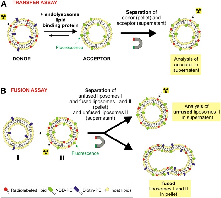
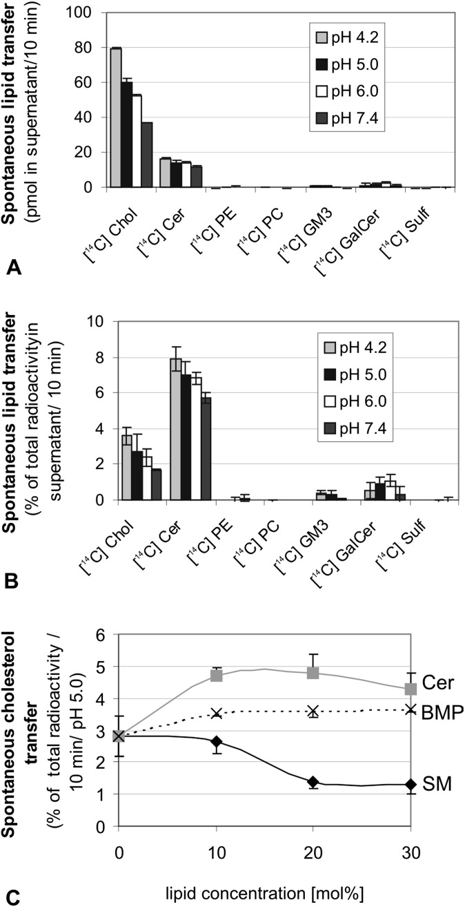
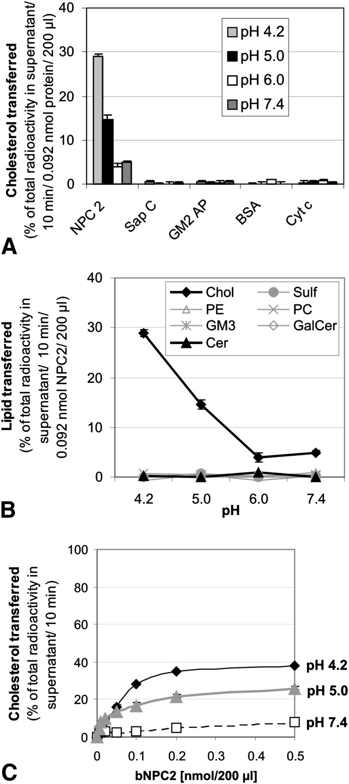
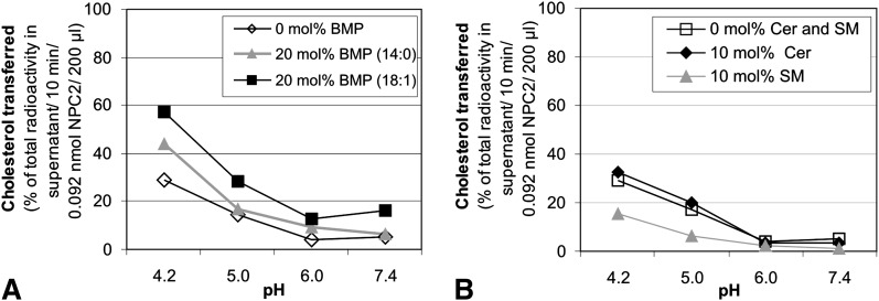
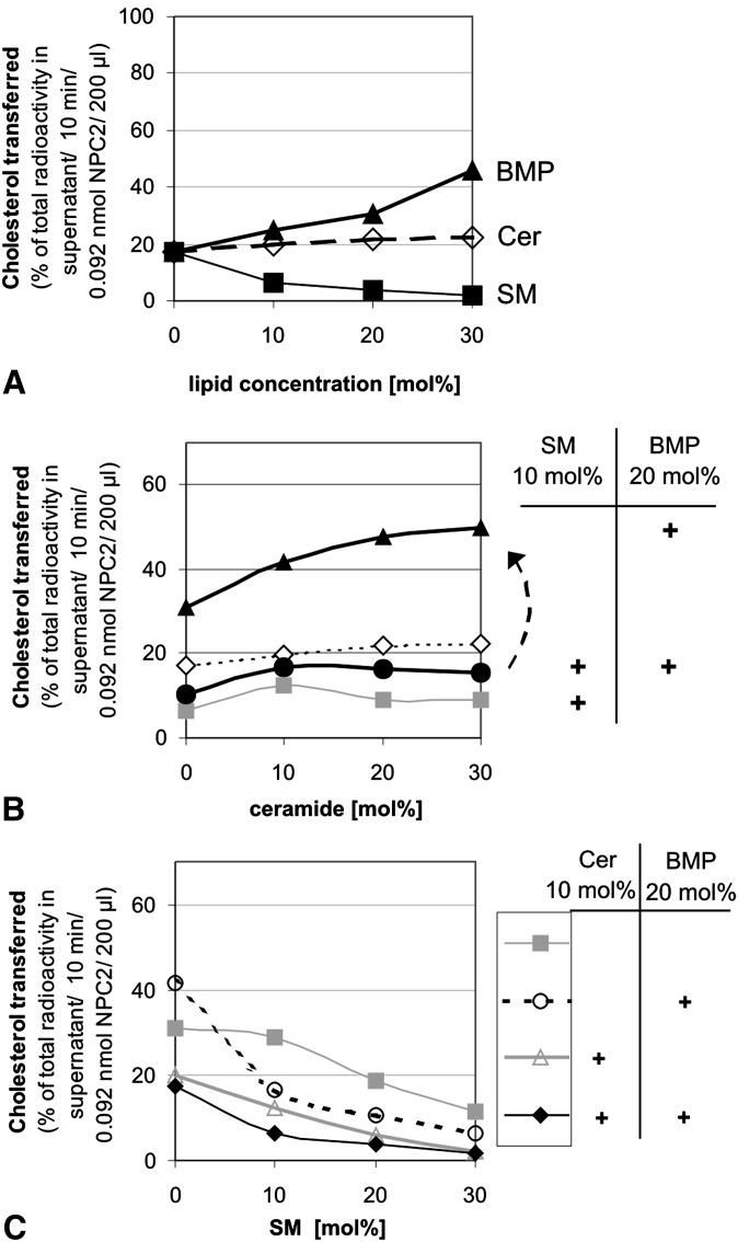
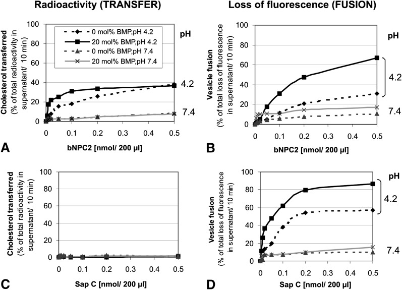
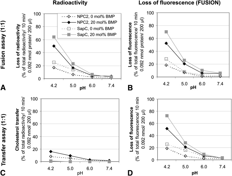
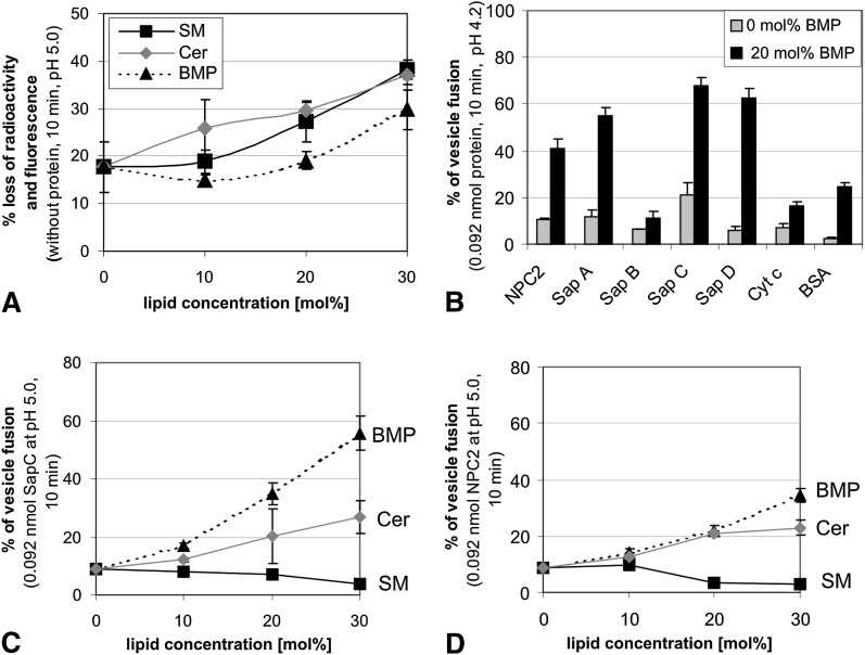
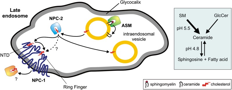
We examined the effect of Niemann-Pick disease type 2 (NPC2) protein and some late endosomal lipids [sphingomyelin, ceramide and bis(monoacylglycero)phosphate (BMP)] on cholesterol transfer and membrane fusion. Of all lipid-binding proteins tested, only NPC2 transferred cholesterol at a substantial rate, with no transfer of ceramide, GM3, galactosylceramide, sulfatide, phosphatidylethanolamine, or phosphatidylserine. Cholesterol transfer was greatly stimulated by BMP, little by ceramide, and strongly inhibited by sphingomyelin. Cholesterol and ceramide were also significantly transferred in the absence of protein. This spontaneous transfer of cholesterol was greatly enhanced by ceramide, slightly by BMP, and strongly inhibited by sphingomyelin. In our transfer assay, biotinylated donor liposomes were separated from fluorescent acceptor liposomes by streptavidin-coated magnetic beads. Thus, the loss of fluorescence indicated membrane fusion. Ceramide induced spontaneous fusion of lipid vesicles even at very low concentrations, while BMP and sphingomyelin did so at about 20 mol% and 10 mol% concentrations, respectively. In addition to transfer of cholesterol, NPC2 induced membrane fusion, although less than saposin-C. In this process, BMP and ceramide had a strong and mild stimulating effect, and sphingomyelin an inhibiting effect, respectively. Note that the effects of the lipids on cholesterol transfer mediated by NPC2 were similar to their effect on membrane fusion induced by NPC2 and saposin-C.


Figures

Fig. 1.
Fig. 1.
Schematic diagram for transfer and fusion assay. A: Transfer assay as described (29) based on the use of donor liposomes containing [14C]labeled lipids and a biotin-labeled phosphatidylethanolamine (PE), which allows the separation of these membranes by streptavidin-coated magnetic beads. The acceptor liposomes are marked by fluorescent NBD-PE to control the recovery of these vesicles. After separation of the biotin-labeled liposomes, we measured radioactivity and fluorescence in the supernatant. B: In another in vitro assay, fusion of liposomes was investigated. One kind of liposomes contained NBD-PE and a radiolabeled lipid (II). If these liposomes (II) fused with Biotin-PE labeled liposomes (I), they would be pulled out by streptavidin-coated magnetic beads together with remaining vesicles (I). So we simultaneously measured a decrease of radioactivity and fluorescence in the supernatant. Red: [14C]labeled lipid e.g., cholesterol; green: NBD-PE; blue: Biotin-PE; yellow: PC as a host lipid.
Fig. 2.
Fig. 2.
Spontaneous transfer of cholesterol and ceramide. Spontaneous transfer (defined as the gain of radiolabeled lipid by acceptor liposomes in the supernatant in the absence of added lysosomal lipid-binding proteins) of different [14C]labeled lipids was determined at different pH values for an incubation time of 10 min at 25°C. The complete amount of spontaneously transferred lipid at different pH values (A) and corresponding chart in percentage of transferred radiolabeled lipid (B). Lipid composition of donor liposome: 10 mol% unlabeled cholesterol, 1 mol% [14C]labeled lipid, 4 mol% Biotin-PE. and 85 mol% PC. The acceptor vesicles contained 10 mol% unlabeled cholesterol, 4 mol% NBD-PE and 86 mol% PC. Spontaneous cholesterol transfer (C) depending on the lipid composition (varying Cer, BMP, and SM content) at pH 5.0 (at pH 4.2 and pH 6.0, similar transfer rates were obtained). Liposomes contained 10 mol% unlabeled cholesterol, 1 mol% [14C] cholesterol, 4 mol% marker lipids, varying Cer, SM or BMP content as given, PC added to 100 mol%. Mean ± SEM (n = 4). Biotin-PE, N-[6-(biotinoyl)amino]hexanoyl-1,2-dihexadecanoyl-sn-glycerol-3-phosphoethanolamine; BMP, bis(monoacylglycero)phosphate; Cer, ceramide; Chol, cholesterol; GalCer, galactosylceramide; NBD-PE, N-(7-nitrobenz-2-oxa-1,3-diazol-4-yl)-1,2-dihexadecanoyl-sn-glycero-3-phosphoethanolamine; NPC2, Niemann-Pick disease type 2 protein; PC, dioleoyl-L-α-phosphatidycholine; PE, phosphatidylethanolamine; SM, sphingomyelin; Sulf, sulfatide.
Fig. 3.
Fig. 3.
Bovine NPC2 transfers [14C] cholesterol under different experimental conditions: (A) cholesterol transfer by different lysosomal proteins, (B) NPC2 specificity for cholesterol transfer, and (C) variations of concentration of bNPC2 at pH 4.2 and 7.4. Donor liposomes contained 10 mol% unlabeled cholesterol, 1 mol% [14C] labeled lipid, 4 mol% Biotin-PE, and 85 mol% PC; and the acceptor liposomes contained 10 mol% unlabeled cholesterol, 4 mol% NBD-PE, and 86 mol% PC. Mean ± SEM (n = 4); in (C), SEM <10% (error bars are within the used symbol). Biotin-PE, N-[6-(biotinoyl)amino]hexanoyl-1,2-dihexadecanoyl-sn-glycerol-3-phosphoethanolamine; BMP, bis(monoacylglycero)phosphate; Cer, ceramide; Chol, cholesterol; cyt C, cytochrom C; GalCer, galactosylceramide; GM2AP, GM2 activator protein; NBD-PE, N-(7-nitrobenz-2-oxa-1,3-diazol-4-yl)-1,2-dihexadecanoyl-sn-glycero-3-phosphoethanolamine; NPC2, Niemann-Pick disease type 2 protein; PC, dioleoyl-L-α-phosphatidycholine; PE, phosphatidylethanolamine; Sap C, saposin C; Sulf, sulfatide.
Fig. 4.
Fig. 4.
pH dependence of cholesterol transfer by NPC2 in the presence of different lipids. A: ± 20 mol% BMP with different chain length. B: ± 10 mol% Cer or ± 10 mol% SM. The lipid composition is the same as in Fig. 2. The supplementation of liposomes with BMP, Cer, or SM is on the expense of the host lipid PC. After 10 min incubation time in the presence of 0.092 nmol bNPC2, the donors were separated by BioMag beads, and the radioactivity and fluorescence of the supernatant were measured. Mean ± SEM in the range of 10% (n = 4) (error bars are within the used symbol). BMP, bis(monoacylglycero)phosphate; Cer, ceramide; NPC2, Niemann-Pick disease type 2 protein; PC, dioleoyl-L-α-phosphatidycholine; SM, sphingomyelin.
Fig. 5.
Fig. 5.
BMP and ceramide stimulate whereas sphingomyelin inhibits bNPC-2–mediated cholesterol transfer at pH 5. A: Increasing concentrations of BMP, Cer, and SM integrated in both donor and acceptor liposomes at a concentration ranging 0–30 mol% on the expense of the host lipid PC. B, C: Combined effects of different lipids on the NPC2 enhanced cholesterol transfer in the presence of increasing Cer (B) and SM concentrations (C). The broken arrow in B points to the increase of cholesterol transfer rate when SM was absent from vesicle membranes. Mean ± SEM in the range of 10% (n = 4) (error bars are within the used symbols). BMP, bis(monoacylglycero)phosphate; Cer, ceramide; NPC2, Niemann-Pick disease type 2 protein; PC, dioleoyl-L-α-phosphatidycholine; SM, sphingomyelin.
Fig. 6.
Fig. 6.
Transfer of [14C] cholesterol and loss of acceptor liposomes (loss of fluorescence) mediated by NPC2 and fusogenic Sap C. A: Cholesterol transfer depending on the concentration of bNPC2 in the presence or absence of 20 mol% BMP. B: bNPC2-mediated fluorescence loss, which implies membrane fusion. As a control we investigated radiolabeled cholesterol transfer (C) and the loss of acceptor (D) mediated by the fusogenic Sap C, containing a C-terminal hexahistidine tag. Mean ± SEM in the range of 12% (n = 4). BMP, bis(monoacylglycero)phosphate; NPC2, Niemann-Pick disease type 2 protein; Sap C, saposin C.
Fig. 7.
Fig. 7.
Fusion assay of liposomes type I with type II. Liposomes I contained Biotin-PE for separation with strepavidin coated magnetic beads. Liposomes II were composed of fluorescent NBD-PE and radiolabeled cholesterol as marker lipids. The ratio of liposomes I and II was 1:1. In case of vesicle fusion, radioactivity (A) and fluorescence (B) should disappear in parallel. A, B: The pH dependence of fusion rate mediated by bNPC2 and Sap C (containing a C-terminal hexahistidine tag) in the presence or absence of 20 mol% BMP. C, D: The corresponding transfer assay with the donor acceptor ratio of 1:1 instead of a ratio of 1:5 as used before. In this transfer assay, radiolabeled cholesterol is in the donor vesicle, and the fluorescent marker lipid NBD-PE is in the acceptor vesicle. Cholesterol transfer is depicted in C and loss of fluorescence in D. Mean ± SEM in the range of 10% (n = 4). BMP, bis(monoacylglycero)phosphate; NPC2, Niemann-Pick disease type 2 protein; Sap C, saposin C.
Fig. 8.
Fig. 8.
Fusion assay measurements. A: Spontaneous fusion in the presence of varying concentrations of SM, Cer, and BMP at pH 5.0. B: Fusion mediated by various endosomal/lysosomal proteins in the presence or absence of 20 mol% BMP at pH 4.2. C, D: Effects of various endosomal lipids on fusion mediated by 0.092 nmol each of hexahistidine tag containing Sap C (C) or bNPC2 (D) at pH 5.0. The ratio of liposome I and II is 1:1. Mean ± SEM (n = 4). BMP, bis(monoacylglycero)phosphate; Cer, ceramide; Cyt c, cytochrom C; NPC2, Niemann-Pick disease type 2 protein; Sap A, saposin A; Sap B, saposin B; Sap C, saposin C; Sap D, saposin D; SM, sphingomyelin.
Fig. 9.
Fig. 9.
Model for lipid sorting at the stage of late endosomes (pH 5.0). Acid sphingomyelinase degrades sphingomyelin to ceramide at the surface of intraendosomal vesicles. Decrease of sphingomyelin and increase of ceramide enhances the availability of cholesterol on inner membranes for NPC2 which removes cholesterol from the inner endosomal vesicles (yellow) and transfers it to the N-terminal domain of NPC1 in the limiting membrane of late endosomes (25). NPC1 mediates cholesterol exit through the glycocalix. ASM, acid sphingomyelinase; NPC, Niemann-Pick type C protein, NTD, N-terminal domain.

Similar articles

Cited by

References

    1. Brown M. S., Goldstein J. L. 1986. A receptor-mediated pathway for cholesterol homeostasis. Science. 232: 34–47. - PubMed
    1. Ikonen E. 2008. Cellular cholesterol trafficking and compartmentalization. Nat. Rev. Mol. Cell Biol. 9: 125–138. - PubMed
    1. Möbius W., van Donselaar E., Ohno-Iwashita Y., Shimada Y., Heijnen H. F., Slot J. W., Geuze H. J. 2003. Recycling compartments and the internal vesicles of multivesicular bodies harbor most of the cholesterol found in the endocytic pathway. Traffic. 4: 222–231. - PubMed
    1. Brown M. S., Goldstein J. L. 1999. A proteolytic pathway that controls the cholesterol content of membranes, cells, and blood. Proc. Natl. Acad. Sci. USA. 96: 11041–11048. - PMC - PubMed
    1. Pentchev P. G., Comly M. E., Kruth H. S., Tokoro T., Butler J., Sokol J., Filling-Katz M., Quirk J. M., Marshall D. C., Patel S., et al. 1987. Group C Niemann-Pick disease: faulty regulation of low-density lipoprotein uptake and cholesterol storage in cultured fibroblasts. FASEB J. 1: 40–45. - PubMed

Publication types

MeSH terms