Comparative Study
. 2010 Mar;66(Pt 3):233-42.
doi: 10.1107/S0907444909054298. Epub 2010 Feb 12.

Structure of the unbound form of HIV-1 subtype A protease: comparison with unbound forms of proteases from other HIV subtypes

Affiliations
Comparative Study

Structure of the unbound form of HIV-1 subtype A protease: comparison with unbound forms of proteases from other HIV subtypes

Arthur H Robbins et al. Acta Crystallogr D Biol Crystallogr. 2010 Mar.

Abstract

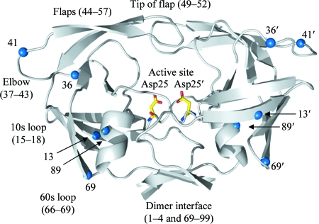
The crystal structure of the unbound form of HIV-1 subtype A protease (PR) has been determined to 1.7 A resolution and refined as a homodimer in the hexagonal space group P6(1) to an R(cryst) of 20.5%. The structure is similar in overall shape and fold to the previously determined subtype B, C and F PRs. The major differences lie in the conformation of the flap region. The flaps in the crystal structures of the unbound subtype B and C PRs, which were crystallized in tetragonal space groups, are either semi-open or wide open. In the present structure of subtype A PR the flaps are found in the closed position, a conformation that would be more anticipated in the structure of HIV protease complexed with an inhibitor. The amino-acid differences between the subtypes and their respective crystal space groups are discussed in terms of the differences in the flap conformations.


Figures

Figure 1
Figure 1
Ribbon diagram of the homodimer of the HIV-1 subtype A PR structure. Catalytic aspartic acids are shown as sticks. Blue spheres indicate amino acids that vary between subtype A PR and subtype B PR (numbers refer to amino-acid positions). Features of the PR structure are as labeled. This figure was generated using PyMOL (DeLano, 2002 ▶).
Figure 2
Figure 2
Stereo 2|F o| − |F c| OMIT map centered on the active site of HIV-1 subtype A PR. The map is contoured at 1.3σ and includes a 4 Å radius buffer around flap residues and water positions. All atoms of the flap (residues 46–55 and 46′–55′), the active-site aspartates Asp25 and Asp25′ and two waters (depicted as red spheres) were omitted from the calculation of the map.
Figure 3
Figure 3
Stereo superposition of unbound PR homodimers. (a) Complete PR homodimers, (b) close-up of the flap regions shown in (a) viewed down the twofold axis. Color code: green, dimers in tetragonal space groups with semi-open flaps; red, dimers in tetragonal space groups with wide-open flaps; blue, dimers in the hexagonal space group P61 with closed flaps. Note that the tips of the flaps invert between the semi-open and closed conformations. PDB coordinates were as used in Fig. 5 ▶. This figure was generated using PyMOL (DeLano, 2002 ▶).
Figure 4
Figure 4
Sequences of HIV-1 PR structures.
Figure 5
Figure 5
Pairwise r.m.s.d.s of Cα—Cα distances (Å).
Figure 6
Figure 6
Stereo figure showing the crystal contacts of HIV-1 subtypes. (a) Wide open (PDB code 2pc0; Heaslet et al., 2007 ▶), (b) semi-open (PDB code 2hb2; Heaslet et al., 2007 ▶) and (c) closed (PDB code 3ixo). PDB coordinates were as used in Fig. 7 ▶. Colored spheres represent the amino acids involved in contacts. A contact is calculated from the sum of atomic radii. This figure was generated using PyMOL (DeLano, 2002 ▶).
Figure 7
Figure 7
Packing contacts (colored cells represent crystal-packing contacts).

Similar articles

Cited by

References

    1. Abecasis, A. B., Deforche, K., Bacheler, L. T., McKenna, P., Carvalho, A. P., Gomes, P., Vandamme, A. M. & Camacho, R. (2006). J. Antivir. Ther.11, 581–589. - PubMed
    1. Baldanti, F., Paolucci, S., Ravasi, G., Maccabruni, A., Moriggia, A., Barbarini, G. & Maserati, R. (2008). J. Med. Virol.80, 947–952. - PubMed
    1. Berman, H. M., Westbrook, J., Feng, Z., Gilliland, G., Bhat, T. N., Weissig, H., Shindyalov, I. N. & Bourne, P. E. (2000). Nucleic Acids Res.28, 235–242. - PMC - PubMed
    1. Brodine, S. K., Mascola, J. R., Weiss, P. J., Ito, S. I., Porter, K. R., Artenstein, A. W., Garland, F. C., McCutchan, F. E. & Burke, D. S. (1995). Lancet, 346, 1198–1199. - PubMed
    1. Brünger, A. T., Adams, P. D., Clore, G. M., DeLano, W. L., Gros, P., Grosse-Kunstleve, R. W., Jiang, J.-S., Kuszewski, J., Nilges, M., Pannu, N. S., Read, R. J., Rice, L. M., Simonson, T. & Warren, G. L. (1998). Acta Cryst. D54, 905–921. - PubMed

Publication types