. 2010 Mar;66(Pt 3):314-8.
doi: 10.1107/S0907444909054535. Epub 2010 Feb 12.

Accommodation of structural rearrangements in the huntingtin-interacting protein 1 coiled-coil domain

Affiliations

Accommodation of structural rearrangements in the huntingtin-interacting protein 1 coiled-coil domain

Jeremy D Wilbur et al. Acta Crystallogr D Biol Crystallogr. 2010 Mar.

Abstract

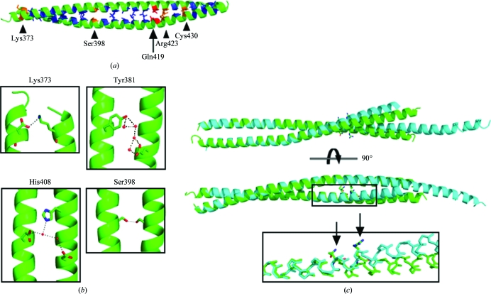
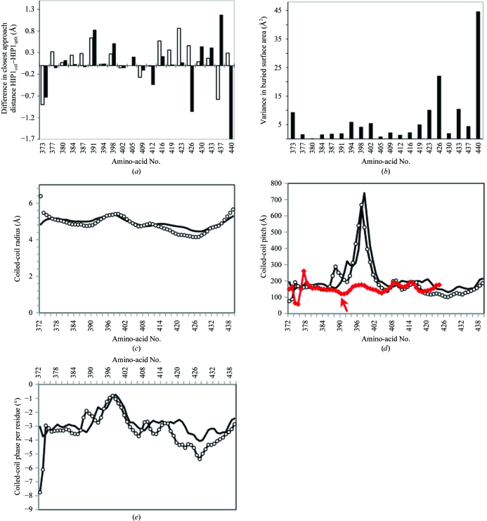
Huntingtin-interacting protein 1 (HIP1) is an important link between the actin cytoskeleton and clathrin-mediated endocytosis machinery. HIP1 has also been implicated in the pathogenesis of Huntington's disease. The binding of HIP1 to actin is regulated through an interaction with clathrin light chain. Clathrin light chain binds to a flexible coiled-coil domain in HIP1 and induces a compact state that is refractory to actin binding. To understand the mechanism of this conformational regulation, a high-resolution crystal structure of a stable fragment from the HIP1 coiled-coil domain was determined. The flexibility of the HIP1 coiled-coil region was evident from its variation from a previously determined structure of a similar region. A hydrogen-bond network and changes in coiled-coil monomer interaction suggest that the HIP1 coiled-coil domain is uniquely suited to allow conformational flexibility.


Figures

Figure 1
Figure 1
Comparison of HIP1 coiled-coil structures. (a) The HIP1 coiled-coil structure determined in this work (residues 367–445; HIP1coil) is shown in green, with core amino acids in other colors. Hydrophobic residues are shown in blue and polar amino acids are shown in orange and red, where those in red have alternate packing in HIP1coil and HIP1split according to SOCKET. (b) Polar contacts (shown as black dashes) found in HIP1coil. The name of one amino acid involved in the polar contact is indicated for reference. (c) Overlay of HIP1coil, shown in green, and the previously determined structure of HIP1 (residues 371–472; HIP1split; PDB code 2qa7; Niu & Ybe, 2008 ▶), shown in cyan. Residues 371–425 were used for alignment. The inset shows the glutamine and arginine residues that occur in the core of the coiled coil and the point of deviation between the two determined structures. Arrows point to the glutamine and arginine side chains. The side chains of other residues have been removed for clarity.
Figure 2
Figure 2
Variations in core-residue packing between HIP1 coiled-coil structures. (a) The distance between the Cα atoms of corresponding residues within each HIP1 coiled-coil homodimer was measured and the difference between the structures was plotted (white bars). The same procedure was used for the difference in closest distances between corresponding side chains within each structure (black bars). (b) The buried surface area for each of the heptad core residues was calculated (each residue occurs four times: twice in each structure). The variance of the mean was plotted for each heptad core residue. (c) Coiled-coil radius, (d) coil pitch and (e) coiled-coil phase per residue relative to residue number as determined using TWISTER (Strelkov & Burkhard, 2002 ▶). HIP1coil, solid black line; HIP1split, open circles; the GIT1 coiled-coil structure (which contains a hydrogen-bonded serine in the coiled-coil core; PDB code 2w6a), red line. GIT1 residue numbering is shifted by −55 for representation on the plot; the arrow points to Ser445.

Similar articles

Cited by

References

    1. Akey, D. L., Malashkevich, V. N. & Kim, P. S. (2001). Biochemistry, 40, 6352–6360. - PubMed
    1. Burkhard, P., Kammerer, R. A., Steinmetz, M. O., Bourenkov, G. P. & Aebi, U. (2000). Structure, 8, 223–230. - PubMed
    1. Chen, C. Y. & Brodsky, F. M. (2005). J. Biol. Chem.280, 6109–6117. - PubMed
    1. Conway, J. F. & Parry, D. A. (1990). Int. J. Biol. Macromol.12, 328–334. - PubMed
    1. Drubin, D. G., Kaksonen, M., Toret, C. & Sun, Y. (2005). Novartis Found. Symp.269, 35–42. - PubMed

Publication types

MeSH terms

Substances