Comment
. 2010 Mar;120(3):660-3.
doi: 10.1172/JCI42378. Epub 2010 Feb 22.

Lights on for aminopeptidases in cystic kidney disease

Affiliations
Comment

Lights on for aminopeptidases in cystic kidney disease

Erwin P Böttinger. J Clin Invest. 2010 Mar.

Abstract

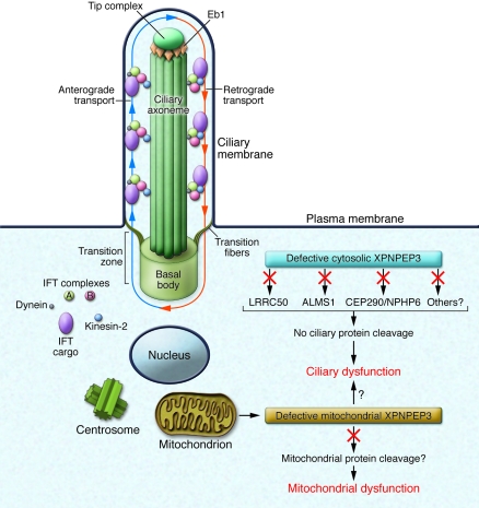
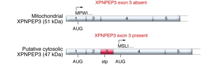
While erudite cell biologists have for many decades described singular immotile appendages known as primary cilia to be present on most cells in our bodies, cilial function(s) long remained an enigma. Driven largely by an ever increasing number of discoveries of genetic defects in primary cilia during the past decade, cilia were catapulted from a long lasting existence in obscurity into the bright spotlight in cell biology and medicine. The study by O'Toole et al. in this issue of the JCI adds a novel "enzymatic" facet to the rapidly growing information about these little cellular tails, by demonstrating that defects in the XPNPEP3 gene, which encodes mitochondrial and cytosolic splice variants of X-prolyl aminopeptidase 3, can cause nephronophthisis-like ciliopathy. Future studies are in order now to elucidate the cystogenic pathways affected by disrupted enzymatic function of XPNPEP3 in cilia-related cystogenic diseases.


Figures

Figure 1
Figure 1. Ciliary and/or mitochondrial dysfunction as consequences of defective ciliary and mitochondrial XPNPEP3.
Cilia consist of a microtubule-based axoneme enveloped by a specialized plasma membrane. The basal body is a microtubule-organizing center that anchors the axoneme. The transition zone (composed of transition fibers) filters molecules that enter or exit the cilium at the junction of the basal body. Axonemal and membrane components are transported in raft macromolecular particles (complexes A and B) by intraflagellar transport (IFT) along the axonemal doublet microtubules toward the tip complex, supported by microtubule end binding protein 1 (Eb1). Anterograde IFT toward the cilial tip is driven by heterotrimeric kinesin-2. Retrograde IFT back to the cell body occurs via the cytoplasmic motor protein dynein. As suggested in the study by O’Toole et al. in this issue of the JCI (13), a cytosolic 48-kDa form of XPNPEP3 may mediate enzymatic cleavage of known cystic-disease proteins ALMS1, LRRC50, and CEP290/NPHP6, and/or other yet unknown ciliary proteins, possibly regulating ciliary targeting or transport. O’Toole et al. report that loss of substrate cleavage, which is associated with defective XPNPEP3, leads to ciliary dysfunction. Similarly, loss of mitochondrial 51-kDa XPNPEP3 peptidase activity may result in failure to cleave yet unknown mitochondrial XPNPEP3 substrates and mitochondrial dysfunction. However, it is unclear at this time whether or how mitochondrial XPNPEP3 defects affect ciliary function. Image adapted with permission from Journal of the American Society of Nephrology (9).
Figure 2
Figure 2. Two alternative splice forms of XPNPEP3 exist in the kidney.
If exon 3 of XPNPEP3 is absent, the XPNPEP3 gene product starts with amino acid sequence MPWI and contains an N-terminal MTS that targets the protein to mitochondria. Exon 3 contains a stop codon (stp) in-frame, resulting in premature termination and removal of the MTS-containing gene product. The alternative XPNPEP3, lacking MTS, is translated from exon 4 AUG, possesses the starting amino acid sequence MSLI, and is likely localized to the cytosol. Mitochondrial XPNPEP3, i.e., the exon 3–spliced form, was used in the localization studies reported in this issue by O’Toole et al. (13).

Comment on

  • Individuals with mutations in XPNPEP3, which encodes a mitochondrial protein, develop a nephronophthisis-like nephropathy.
    O'Toole JF, Liu Y, Davis EE, Westlake CJ, Attanasio M, Otto EA, Seelow D, Nurnberg G, Becker C, Nuutinen M, Kärppä M, Ignatius J, Uusimaa J, Pakanen S, Jaakkola E, van den Heuvel LP, Fehrenbach H, Wiggins R, Goyal M, Zhou W, Wolf MT, Wise E, Helou J, Allen SJ, Murga-Zamalloa CA, Ashraf S, Chaki M, Heeringa S, Chernin G, Hoskins BE, Chaib H, Gleeson J, Kusakabe T, Suzuki T, Isaac RE, Quarmby LM, Tennant B, Fujioka H, Tuominen H, Hassinen I, Lohi H, van Houten JL, Rotig A, Sayer JA, Rolinski B, Freisinger P, Madhavan SM, Herzer M, Madignier F, Prokisch H, Nurnberg P, Jackson PK, Khanna H, Katsanis N, Hildebrandt F. O'Toole JF, et al. J Clin Invest. 2010 Mar;120(3):791-802. doi: 10.1172/JCI40076. Epub 2010 Feb 22. J Clin Invest. 2010. PMID: 20179356 Free PMC article.

Similar articles

Cited by

References

    1. Pazour GJ, Witman GB. The vertebrate primary cilium is a sensory organelle. Curr Opin Cell Biol. 2003;15(1):105–110. doi: 10.1016/S0955-0674(02)00012-1. - DOI - PubMed
    1. Gerdes JM, Davis EE, Katsanis N. The vertebrate primary cilium in development, homeostasis, and disease. Cell. 2009;137(1):32–45. doi: 10.1016/j.cell.2009.03.023. - DOI - PMC - PubMed
    1. Singla V, Reiter JF. The primary cilium as the cell’s antenna: signaling at a sensory organelle. Science. 2006;313(5787):629–633. doi: 10.1126/science.1124534. - DOI - PubMed
    1. Tobin JL, Beales PL. The nonmotile ciliopathies. Genet Med. 2009;11(6):386–402. doi: 10.1097/GIM.0b013e3181a02882. - DOI - PubMed
    1. Scholey JM. Intraflagellar transport motors in cilia: moving along the cell’s antenna. J Cell Biol. 2008;180(1):23–29. doi: 10.1083/jcb.200709133. - DOI - PMC - PubMed

Publication types

MeSH terms