. 2010 Mar;120(3):924-30.
doi: 10.1172/JCI40094. Epub 2010 Feb 22.

Human liver chimeric mice provide a model for hepatitis B and C virus infection and treatment

Affiliations

Human liver chimeric mice provide a model for hepatitis B and C virus infection and treatment

Karl-Dimiter Bissig et al. J Clin Invest. 2010 Mar.

Abstract

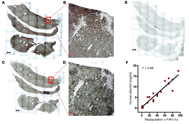
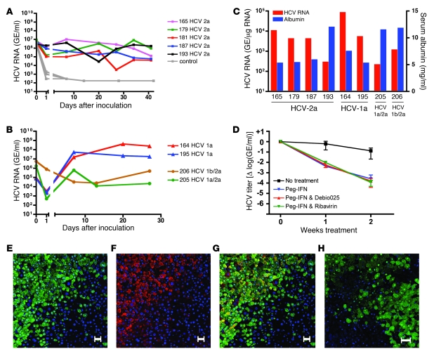
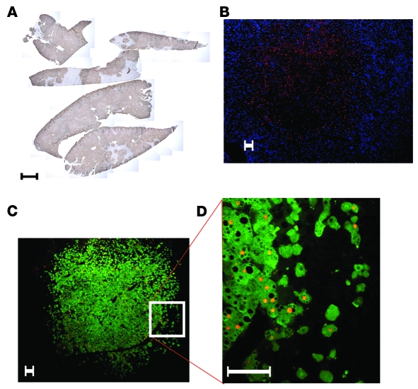
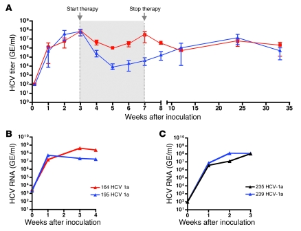
A paucity of versatile small animal models of hepatitis B virus (HBV) and hepatitis C virus (HCV) infection has been an impediment to both furthering understanding of virus biology and testing antiviral therapies. We recently described a regulatable system for repopulating the liver of immunodeficient mice (specifically mice lacking fumaryl acetoacetate hydrolase [Fah], recombination activating gene 2 [Rag2], and the gamma-chain of the receptor for IL-2 [Il-2rgamma]) with human hepatocytes. Here we have shown that a high transplantation dose (3 x 10⁶ to 5 x 10⁶ human hepatocytes/mouse) generates a higher rate of liver chimerism than was previously obtained in these mice, up to 95% human hepatocyte chimerism. Mice with a high level of human liver chimerism propagated both HBV and HCV, and the HCV-infected mice were responsive to antiviral treatment. This human liver chimeric mouse model will expand the experimental possibilities for studying HBV and HCV infection, and possibly other human hepatotropic pathogens, and prove useful for antiviral drug testing.


Figures

Figure 1
Figure 1. Nearly complete repopulation of the Fah–/–Rag2–/–Il2rg–/– mouse liver with human hepatocytes.
(AE) Immunostaining of the same liver for FAH (A and B), human-specific cytokeratin-18 (C and D), and no primary antibody (control, E). (B and D) Higher-magnification views of the boxed areas in A and C, respectively. Scale bars: 1 mm (A, C, and E), 50 μm (B and D). (F) Correlation of human albumin level in the murine serum and repopulation as assessed by morphometry of FAH immunostaining. A, C, and E are composite images.
Figure 3
Figure 3. HCV infection of mice with high human chimerism.
HCV RNA in mouse serum after inoculation with HCV genotype 2a (A) or clinical isolate of HCV genotype 1a or chimeric HCV genotypes (1a/2a and 1b/2a) (B). (C) HCV RNA intermediates detected in chimeric livers and human albumin levels in the murine serum of HCV-infected mice. HCV RNA was normalized to human GAPDH levels and expressed as GE per μg total RNA. (D) Antiviral treatment with peg-IFN alone, peg-IFN and ribavirin, and peg-IFN and Debio 025 for 2 weeks. Results are shown as normalized mean ± SD. (EG) Fluorescent costaining of an HCV-infected chimeric liver for FAH (green) and HCV NS5a (red); nuclear counterstaining with DAPI (blue). (E) FAH and nuclear staining. (F) NS5a and nuclear staining. (G) Merge of E and F showing colocalization of HCV and human hepatocytes in yellow. (H) Control mouse liver repopulated with human hepatocytes but not inoculated with HCV. Costaining for FAH (green) and HCV NS5a (red); nuclear counterstaining with DAPI (blue). Scale bars: 50 μm.
Figure 2
Figure 2. HBV-infected mouse with high human chimerism.
(A) Immuno­staining for FAH and counterstaining with hematoxylin shows that 80% of the liver consists of human hepatocytes. (B) Immunostaining for HBcAg (red) and counterstaining with DAPI (blue). (C and D) Fluorescent costaining for FAH (green) and HBcAg (red). The image in D is a higher-magnification view of the boxed area in C. B and C show the same human hepatocyte cluster. Scale bars: 1 mm (A), 50 μm (BD). A is a composite image.
Figure 4
Figure 4. Long-term propagation and serial passage of HCV.
(A) Mice were treated for 4 weeks with peg-IFN and Debio 025. The treatment was started 3 weeks after inoculation with HCV genotype 1a, when the viral titers reached the plateau phase. Serum HCV RNA levels are presented as mean ± range of treatment (blue, n = 3) and control (red, n = 3) groups. (B) Two mice were inoculated with a clinical isolate of HCV genotype 1a. (C) Serum from mouse 164 and 195 was used as inoculum for mouse 235 and 239 (see text for details).

Comment in

Similar articles

Cited by

References

    1. Seeger C, Mason WS. Hepatitis B virus biology. Microbiol Mol Biol Rev. 2000;64(1):51–68. doi: 10.1128/MMBR.64.1.51-68.2000. - DOI - PMC - PubMed
    1. Wieland SF, Chisari FV. Stealth and cunning: hepatitis B and hepatitis C viruses. J Virol. 2005;79(15):9369–9380. - PMC - PubMed
    1. Dienstag JL. Hepatitis B virus infection. N Engl J Med. 2008;359(14):1486–1500. doi: 10.1056/NEJMra0801644. - DOI - PubMed
    1. Llovet JM, Burroughs A, Bruix J. Hepatocellular carcinoma. Lancet. 2003;362(9399):1907–1917. doi: 10.1016/S0140-6736(03)14964-1. - DOI - PubMed
    1. Barker LF, et al. Transmission of type B viral hepatitis to chimpanzees. J Infect Dis. 1973;127(6):648–662. - PubMed

Publication types