. 2010 Jun;18(6):1130-1142.
doi: 10.1038/mt.2010.29. Epub 2010 Feb 23.

Inhibition of multiple protective signaling pathways and Ad.5/3 delivery enhances mda-7/IL-24 therapy of malignant glioma

Affiliations

Inhibition of multiple protective signaling pathways and Ad.5/3 delivery enhances mda-7/IL-24 therapy of malignant glioma

Hossein A Hamed et al. Mol Ther. 2010 Jun.

Retraction in

Abstract

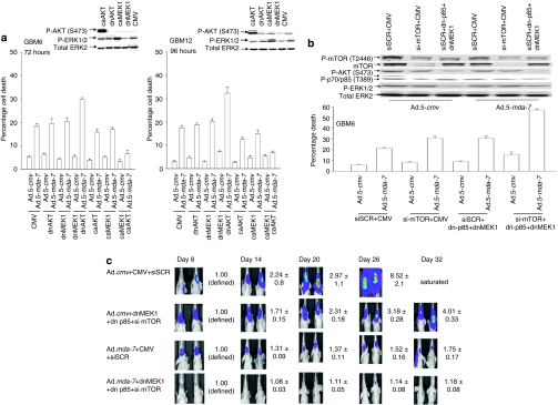
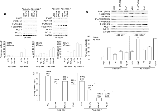
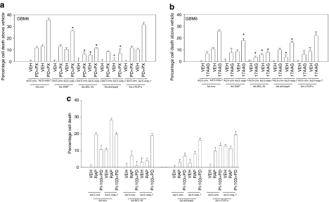
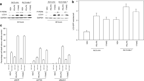
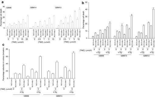
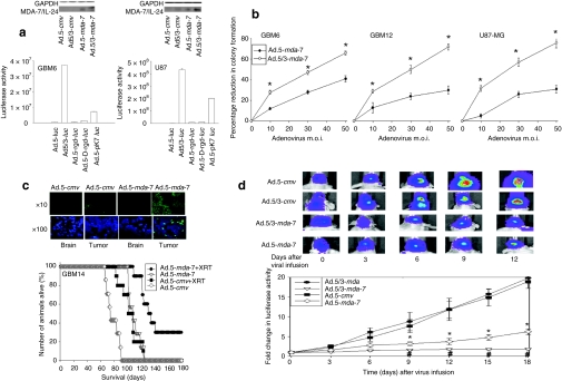
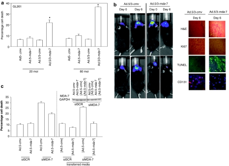
We have explored the mechanism by which inhibition of multiple cytoprotective cell-signaling pathways enhance melanoma differentiation-associated gene-7/interleukin-24 (mda-7/IL-24) toxicity toward invasive primary human glioblastoma multiforme (GBM) cells, and whether improving adenoviral infectivity/delivery of mda-7/IL-24 enhances therapeutic outcome in animals containing orthotopic xenografted GBM cells. The toxicity of a serotype 5 recombinant adenovirus to express MDA-7/IL-24 (Ad.5-mda-7) was enhanced by combined molecular or small molecule inhibition of mitogen-activated extracellular regulated kinase (MEK)1/2 and phosphatidyl inositol 3-kinase (PI3K) or AKT; inhibition of mammalian target of rapamycin (mTOR) and MEK1/2; and the HSP90 inhibitor 17AAG. Molecular inhibition of mTOR/PI3K/MEK1 signaling in vivo also enhanced Ad.5-mda-7 toxicity. In GBM cells of diverse genetic backgrounds, inhibition of cytoprotective cell-signaling pathways enhanced MDA-7/IL-24-induced autophagy, mitochondrial dysfunction and tumor cell death. Due partly to insufficient adenovirus serotype 5 gene delivery this therapeutic approach has shown limited success in GBM. To address this problem, we employed a recombinant adenovirus that comprises the tail and shaft domains of a serotype 5 virus and the knob domain of a serotype 3 virus expressing MDA-7/IL-24, Ad.5/3-mda-7. Ad.5/3-mda-7 more effectively infected and killed GBM cells in vitro and in vivo than Ad.5-mda-7. Future combinations of these approaches hold promise for developing an effective therapy for GBM.


Figures

<b>Figure 1</b>
Figure 1
Ad.5-mda-7 lethality is enhanced by combined inhibition of PI3K/MEK/mTOR pathways. (a) GBM6 and GBM12 cells were infected with empty vector control virus (Ad.5-cmv) or with viruses to express MDA-7/IL-24 (Ad.5-mda-7), activated forms of AKT and MEK1 (caAKT, caMEK1), and dominant negative forms of AKT and MEK1 (dnAKT, dnMEK1). At 48 hours (GBM6) or 96 hours (GBM12) after infection, cells were isolated and cell viability was determined by Trypan blue exclusion assay (±SEM, n = 3). Upper inset shows the impact of expressing caAKT, caMEK1, dnAKT, and dnMEK1 on cell signaling. (b) GBM6 cells were infected with empty vector control virus (Ad.5-cmv) or with a virus to express MDA-7/IL-24 (Ad.5-mda-7). In parallel, cells were transfected with empty vector plasmid or plasmids to express a dn-p85 PI3K subunit (dn-p85) and dnMEK1; cells were transfected with a nonspecific scrambled siRNA (siSCR) or an siRNA to knockdown mTOR expression, an indicated. At 48 hours after infection, cells were isolated and cell viability was determined by Trypan blue exclusion assay (±SEM, n = 3). (c) GBM6-luciferase cells were infected with empty vector control virus (Ad.5-cmv) or with viruses to express MDA-7/IL-24 (Ad.5-mda-7) and in parallel transfected with a siSCR and an empty vector plasmid or with an siRNA to knockdown mTOR expression and plasmids to express a dn-p85 PI3K subunit and dnMEK1. At 12 hours after transfection/infection equal numbers of viable tumor cells were implanted into the brains of athymic mice and tumor formation monitored over the following 32 days by luciferase/CCD camera imaging (n = 2, ±SEM). CMV, cytomegalovirus; dn, dominant negative; ERK, extracellular regulated kinase; GBM, glioblastoma multiforme; MEK, mitogen-activated extracellular regulated kinase.
<b>Figure 2</b>
Figure 2
Inhibition of mTOR, PI3K, and MEK1/2 signaling enhances Ad.5-mda-7 lethality in GBM cells. (a) GBM6, GBM12, and GBM14 cells were infected with empty vector (Ad.5-cmv) or to express MDA-7/IL-24 (Ad.5-mda-7) and 12 hours after infection treated with vehicle (VEH) [dimethyl sulfoxide (DMSO)], PD184352+PX866 (PD+PX, 1 µmol/l + 100 nmol/l), 17AAG (100 nmol/l). At 48 hours after infection, cells were isolated and cell viability was determined by Trypan blue exclusion assay (±SEM, n = 3). Upper blots: GBM6 cells were infected with Ad.5-cmv or Ad.5-mda-7 and 12 hours after infection treated with vehicle (DMSO), 17AAG (100 nmol/l), or PD184352 (PD, 1 µmol/l) and PX866 (100 nmol/l) (PD+PX). At 24 and 48 hours after infection, cells were isolated and sodium dodecyl sulfate–polyacrylamide gel electrophoresis (SDS-PAGE) was performed to determine the expression of BCL-XL and MCL-1, and the phosphorylation of ERK1/2, p38 MAPK, JNK1-3, AKT (S473) (n = 2). (b) GBM6 cells were infected with Ad.5-cmv or Ad.5-mda-7 and 12 hours after infection treated with vehicle (DMSO), rapamycin (rap, 100 nmol/l), PI-103 (200 nmol/l), PD184352 (PD, 1 µmol/l), or PI-103+PD. At 48 hours after infection, cells were isolated and cell viability was determined by Trypan blue exclusion assay (±SEM, n = 3). Upper blots: GBM6 cells were infected with Ad.5-cmv or Ad.5-mda-7 and 12 hours after infection treated with vehicle (DMSO), rapamycin (rap, 100 nmol/l), or PD184352 (PD, 1 µmol/l) and PI-103 (200 nmol/l) (PD+PI-103). At 24hour(s) after infection cells were isolated and SDS-PAGE performed to determine the expression of BCL-XL and MCL-1, and the phosphorylation of ERK1/2, p38 MAPK, JNK1-3, AKT (S473), mTOR (S1221), and p70 S6K (T389) (n = 2). (c) GBM6 cells were plated as single cells in sextuplicate and 12 hours after plating were infected with Ad.5-cmv or Ad.5-mda-7. At 12hour(s) after infection cells were treated with vehicle (DMSO), PD184352 (PD, 1 µmol/l) + PI-103 (200 nmol/l) (PD+PI), PD184352 (PD, 1 µmol/l) + PX866 (100 nmol/l) (PD+PX), or 17AAG (100 nmol/l). At 48 hours after infection, the growth media was removed and replaced with new media lacking drugs. Colonies of >50 cells were permitted to form over the following ~20 days, followed by fixing, staining, and counting (±SEM, n = 3). GBM, glioblastoma multiforme; MEK, mitogen-activated extracellular regulated kinase; mTOR, mammalian target of rapamycin; PI3K, phosphatidyl inositol 3 kinase.
<b>Figure 3</b>
Figure 3
Activation of the intrinsic pathway plays a key role in the facilitation of Ad.5-mda-7 toxicity by kinase inhibitors. (a) GBM6 cells were infected with Ad.5-cmv or Ad.5-mda-7 in combination with viruses to express XIAP, BCL-XL, dominant negative (dn) caspase 9, or c-FLIP-s, and 12 hours after infection treated with vehicle [VEH; dimethyl sulfoxide (DMSO)], or PD184352 (PD, 1 µmol/l) + PX866 (100 nmol/l) (PD+PX). At 48 hours after infection cells were isolated and cell viability was determined by Trypan blue exclusion assay (±SEM, n = 3). (b) GBM6 cells were infected with Ad.5-cmv or Ad.5-mda-7 in combination with viruses to express XIAP, BCL-XL, dn-caspase 9, or c-FLIP-s, and 12 hours after infection treated with VEH (DMSO), or 17AAG (100 nmol/l). At 48 hours after infection cells were isolated and cell viability was determined by Trypan blue exclusion assay (±SEM, n = 3). (c) GBM6 cells were infected with Ad.5-cmv or Ad.5-mda-7 in combination with viruses to express XIAP, BCL-XL, dn-caspase 9, or c-FLIP-s, and 12 hours after infection treated with VEH (DMSO), rapamycin (100 nmol/l), or PI-103 (200 nmol/l) + PD184352 (PD, 1 µmol/l) (PI-103+PD). At 48 hours after infection cells were isolated and cell viability was determined by Trypan blue exclusion assay (±SEM, n = 3). GBM, glioblastoma multiforme.
<b>Figure 4</b>
Figure 4
Inhibition of signaling pathways enhances Ad.5-mda-7-induced autophagy, but does not promote additional activation of PERK. (a) GBM6 cells were infected with Ad.5-cmv or Ad.5-mda-7 and 12 hours after infection treated with vehicle [VEH; dimethyl sulfoxide (DMSO)], PD184352 (PD, 1 µmol/l) + PX866 (100 nmol/l) (PD+PX), or 17AAG (100 nmol/l). Cells were isolated 24 and 48 hours after infection and sodium dodecyl sulfate–polyacrylamide gel electrophoresis and immunoblotting was performed to assess the phosphorylation of PERK and the conversion of LC3 to LC3-II (n = 3). (b) GBM6 cells in 4-well chambered slides were infected with Ad.5-cmv or Ad.5-mda-7 and in parallel transfected with a plasmid to express LC3-GFP and with either a scrambled siRNA (siSCR) or an siRNA to knockdown Beclin1 (siBeclin1). At 12 hours after infection cells were treated with vehicle (DMSO), 17AAG, or PD+PX. At 12 hours after drug treatment cells were examined using a fluorescent microscope (×40 magnification) for the formation of intense GFP-staining vesicles (±SEM, n = 3). (c) GBM6 cells were infected with Ad.5-cmv or Ad.5-mda-7 and in parallel transfected with either a scrambled siRNA (siSCR) or siRNA molecules to knockdown ATG5 (siATG5) or Beclin1 (siBeclin1). At 12 hours after infection cells were treated with vehicle (DMSO), PD184352 (PD, 1 µmol/l) + PX866 (100 nmol/l) (PD+PX), or 17AAG (100 nmol/l). At 48 hours after infection cells were isolated and cell viability was determined by Trypan blue exclusion assay (±SEM, n = 3). GBM, glioblastoma multiforme; PERK, protein kinase R–like endoplasmic reticulum kinase.
<b>Figure 5</b>
Figure 5
Temozolomide (TMZ) potentiates Ad.5-mda-7 lethality. (a) GBM6, GBM12, and GBM14 cells were infected with Ad.5-cmv or Ad.5-mda-7, and 12 hours after infection cells were treated with vehicle [dimethyl sulfoxide (DMSO)] or increasing concentrations of TMZ. At 48 hours after infection, cells were isolated and cell viability was determined by TUNEL assay (±SEM, n = 3). (b) GBM6, GBM12, and GBM14 cells were infected with Ad.5-cmv or Ad.5-mda-7, and 12 hours after infection cells were treated with vehicle (DMSO) or increasing concentrations of TMZ. At 24 hours after virus infection cells were irradiated (4 Gy). At 48 hours after infection, cells were isolated and cell viability was determined by TUNEL assay (±SEM, n = 3). (c) GBM6 and GBM14 cells were plated as single cells and were infected with Ad.5-cmv or Ad.5-mda-7, and 12 hours after infection cells were treated with vehicle (DMSO) or increasing concentrations of TMZ. At 24 hours after virus infection cells were irradiated (4 Gy). At 48 hours after infection, the media was replaced with media lacking vehicle/drugs. Colonies of >50 cells were permitted to form over the following ~20 days, followed by fixing, staining, and counting (±SEM, n = 3). GBM, glioblastoma multiforme.
<b>Figure 6</b>
Figure 6
A tropism-modified type 5/type 3 adenovirus infects GBM cells more readily than a type 5 adenovirus, and enhances tumor killing in vivo. (a) GBM6 (CAR+++) and U87-MG (CAR+) cells were infected with Ad.5-luc or with tropism-modified viruses Ad.5/3-luc, Ad.5-rgd-luc, Ad.5-D-rgd-luc, Ad.5-pK7-luc at 10 MOI, as indicated. Cells were lysed 48 hours after infection and the luciferase activity was measured in triplicate (±SEM, n = 3). Upper blot: U87-MG and GBM6 cells were infected with Ad.5-cmv, Ad.5/3-cmv, Ad.5-mda-7 or Ad.5/3-mda-7 (10 MOI). Cells were isolated 24 hours after infection and MDA-7/IL-24 protein levels determined (n = 2). (b) GBM6, GBM12, and U87-MG cells were plated in sextuplicate as single cells and were infected with Ad.5-cmv or Ad.5-mda-7 or with tropism-modified viruses Ad.5/3-cmv or Ad.5/3-mda-7 at 0–50 multiplicity of infection (MOI). Colonies of >50 cells were permitted to form over 14–20 days, followed by fixing, staining, and counting. Data are presented as the real percentage reduction in colony formation subtracting the effect of Ad.cmv (5 or 5/3) infection (±SEM, n = 3) *P < 0.05 greater than Ad.5-mda-7 infection. (c) GBM14 cells were implanted into the brains of athymic nude mice and 14 days after implantation tumors were infused with Ad.5-cmv or Ad.5-mda-7 and 48 hours after infection the heads of each animal were irradiated (2 × 4 Gy, once every 24 hours). Animals were monitored daily and when approaching death were killed and survival of animals is plotted as a percentage of animals alive on any given day (±SEM, n = 2). Inset panel: 2 days after infection, animal brains were isolated and TUNEL (terminal deoxynucleotidyl transferase dUTP nick-end labeling) stained for apoptotic cells. (d) GBM6 cells that stably express luciferase were implanted into the brains of athymic nude mice and 14 days after implantation tumors were infused with Ad.5-cmv or Ad.5-mda-7, or Ad.5/3-cmv or Ad.5/3-mda-7. The tumors in situ were imaged using a Xenogen CCD system and the light intensity quantified before and for 18 days following adenovirus infusion (±SEM, n = 2). *P < 0.05 less than corresponding Ad.5-cmv treated cells; #P < 0.05 less than Ad.5-mda-7 value. GAPDH, glyceraldehyde 3-phosphate dehydrogenase; GBM, glioblastoma multiforme.
<b>Figure 7</b>
Figure 7
Ad.5/3-mda-7 is more effective at killing and reducing rodent GL261 glioma viability in vitro and in vivo. (a) GL261 cells were infected with Ad.5-cmv or Ad.5-mda-7 or with Ad.5/3-cmv or Ad.5/3-mda-7 at 20 or 80 multiplicity of infection (MOI). At 48 hours after infection, cells were isolated and cell viability was determined by Trypan blue dye exclusion assay (±SEM, n = 3). *P < 0.05 greater than Ad.5-mda-7 value. (b) C57 black mice were implanted with GL261 cells stably expressing luciferase. At 14 days after implantation tumors were infused with Ad.5/3-cmv or Ad.5/3-mda-7. The tumors in situ were imaged using a Xenogen CCD system and the light intensity quantified before and 6 days following adenovirus infusion (n = 2). Inset panel: 6 days after virus infusion mouse brains were isolated and immunohistochemistry performed to detect the levels of Ki67, CD131, and TUNEL (terminal deoxynucleotidyl transferase dUTP nick-end labeling) staining (n = 2). (c) GBM6 cells were infected with Ad.5-cmv or Ad.5-mda-7 and in parallel transfected with plasmids to express a control shRNA (shSCR) or an shRNA to knockdown MDA-7/IL-24 expression (shMDA-7). At 48 hours after infection, cells and media were isolated. Cell viability was determined by Trypan blue dye exclusion assay (±SEM, n = 3). The media was placed onto GBM6 cells that had been transfected with plasmids to express a control shRNA (shSCR) or an shRNA to knockdown MDA-7/IL-24 expression (shMDA-7) (labeled in parentheses, grey). At 48 hours after placement of conditioned media onto the cells, cell viability was again determined by Trypan blue exclusion assay (±SEM, n = 3).

References

    1. Robins HI, Chang S, Butowski N., and , Mehta M. Therapeutic advances for glioblastoma multiforme: current status and future prospects. Curr Oncol Rep. 2007;9:66–70. - PubMed
    1. Jiang H, Lin JJ, Su ZZ, Goldstein NI., and , Fisher PB. Subtraction hybridization identifies a novel melanoma differentiation associated gene, mda-7, modulated during human melanoma differentiation, growth and progression. Oncogene. 1995;11:2477–2486. - PubMed
    1. Ekmekcioglu S, Ellerhorst J, Mhashilkar AM, Sahin AA, Read CM, Prieto VG, et al. Down-regulated melanoma differentiation associated gene (mda-7) expression in human melanomas. Int J Cancer. 2001;94:54–59. - PubMed
    1. Ellerhorst JA, Prieto VG, Ekmekcioglu S, Broemeling L, Yekell S, Chada S, et al. Loss of MDA-7 expression with progression of melanoma. J Clin Oncol. 2002;20:1069–1074. - PubMed
    1. Huang EY, Madireddi MT, Gopalkrishnan RV, Leszczyniecka M, Su Z, Lebedeva IV, et al. Genomic structure, chromosomal localization and expression profile of a novel melanoma differentiation associated (mda-7) gene with cancer specific growth suppressing and apoptosis inducing properties. Oncogene. 2001;20:7051–7063. - PubMed

Publication types