

Low oxygen tension and synthetic nanogratings improve the uniformity and stemness of human mesenchymal stem cell layer
- PMID: 20179678
- PMCID: PMC2890122
- DOI: 10.1038/mt.2010.21
Low oxygen tension and synthetic nanogratings improve the uniformity and stemness of human mesenchymal stem cell layer
Abstract
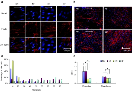
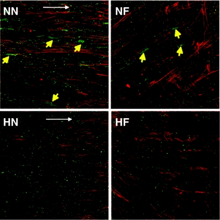
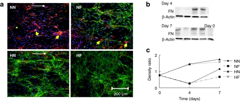
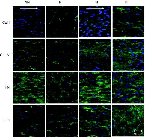
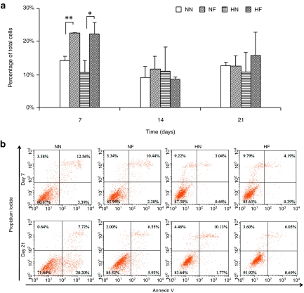
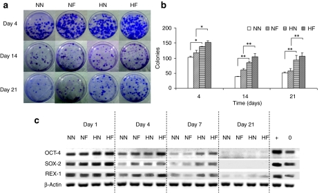
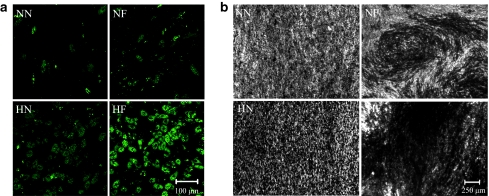
A free-standing, robust cell sheet comprising aligned human mesenchymal stem cells (hMSCs) offers many interesting opportunities for tissue reconstruction. As a first step toward this goal, a confluent, uniform hMSC layer with a high degree of alignment and stemness maintenance needs to be created. Hypothesizing that topographical cue and a physiologically relevant low-oxygen condition could promote the formation of such an hMSC layer, we studied the culture of hMSCs on synthetic nanogratings (350 nm width and 700 nm pitch) and either under 2 or 20% O(2). Culturing hMSCs on the nanogratings highly aligned the cells, but it tended to create patchy layers and accentuate the hMSC differentiation. The 2% O(2) improved the alignment and uniformity of hMSCs, and reduced their differentiation. Over a 14-day culture period, hMSCs in 2% O(2) showed uniform connexon distribution, secreted abundant extracellular matrix (ECM) proteins, and displayed a high progenicity. After 21-day culture on nanogratings, hMSCs exposed to 2% O(2) maintained a higher viability and differentiation capacity. This study established that a 2% O(2) culture condition could restrict the differentiation of hMSCs cultured on nanopatterns, thereby setting the foundation to fabricate a uniformly aligned hMSC sheet for different regenerative medicine applications.
Figures
Similar articles
-
Prevascularization of natural nanofibrous extracellular matrix for engineering completely biological three-dimensional prevascularized tissues for diverse applications.J Tissue Eng Regen Med. 2018 Mar;12(3):e1325-e1336. doi: 10.1002/term.2512. Epub 2017 Nov 27. J Tissue Eng Regen Med. 2018. PMID: 28714140 Free PMC article.
-
Effects of Fiber Alignment and Coculture with Endothelial Cells on Osteogenic Differentiation of Mesenchymal Stromal Cells.Tissue Eng Part C Methods. 2020 Jan;26(1):11-22. doi: 10.1089/ten.TEC.2019.0232. Epub 2019 Dec 27. Tissue Eng Part C Methods. 2020. PMID: 31774033
-
Perfusion affects the tissue developmental patterns of human mesenchymal stem cells in 3D scaffolds.J Cell Physiol. 2009 May;219(2):421-9. doi: 10.1002/jcp.21688. J Cell Physiol. 2009. PMID: 19170078
-
The Role of Dissolved Oxygen Levels on Human Mesenchymal Stem Cell Culture Success, Regulatory Compliance, and Therapeutic Potential.Stem Cells Dev. 2018 Oct 1;27(19):1303-1321. doi: 10.1089/scd.2017.0291. Epub 2018 Aug 22. Stem Cells Dev. 2018. PMID: 30003826 Review.
-
[Safety evaluation of tissue engineered medical devices using normal human mesenchymal stem cells].Yakugaku Zasshi. 2007 May;127(5):851-6. doi: 10.1248/yakushi.127.851. Yakugaku Zasshi. 2007. PMID: 17473528 Review. Japanese.
Cited by
-
Stem cells and cell therapies in lung biology and lung diseases.Proc Am Thorac Soc. 2011 Jun;8(3):223-72. doi: 10.1513/pats.201012-071DW. Proc Am Thorac Soc. 2011. PMID: 21653527 Free PMC article.
-
Nanotopographical control of stem cell differentiation.J Tissue Eng. 2010 Aug 18;2010:120623. doi: 10.4061/2010/120623. J Tissue Eng. 2010. PMID: 21350640 Free PMC article.
-
Direct Control of Stem Cell Behavior Using Biomaterials and Genetic Factors.Stem Cells Int. 2018 May 10;2018:8642989. doi: 10.1155/2018/8642989. eCollection 2018. Stem Cells Int. 2018. PMID: 29861745 Free PMC article. Review.
-
Aligned Nanofibrous Cell-Derived Extracellular Matrix for Anisotropic Vascular Graft Construction.Adv Healthc Mater. 2017 May;6(10):10.1002/adhm.201601333. doi: 10.1002/adhm.201601333. Epub 2017 Feb 9. Adv Healthc Mater. 2017. PMID: 28181412 Free PMC article.
-
Discrepancies on the Role of Oxygen Gradient and Culture Condition on Mesenchymal Stem Cell Fate.Adv Healthc Mater. 2021 Mar;10(6):e2002058. doi: 10.1002/adhm.202002058. Epub 2021 Feb 2. Adv Healthc Mater. 2021. PMID: 33533187 Free PMC article. Review.
References
-
- Pittenger MF, Mackay AM, Beck SC, Jaiswal RK, Douglas R, Mosca JD, et al. Multilineage potential of adult human mesenchymal stem cells. Science. 1999;284:143–147. - PubMed
-
- Chen J, Zhang ZG, Li Y, Wang L, Xu YX, Gautam SC, et al. Intravenous administration of human bone marrow stromal cells induces angiogenesis in the ischemic boundary zone after stroke in rats. Circ Res. 2003;92:692–699. - PubMed
-
- Horwitz EM, Prockop DJ, Fitzpatripnck LA, Koo WW, Gordon PL, Neel M, et al. Transplantability and therapeutic effects of bone marrow-derived mesenchymal cells in children with osteogenesis imperfecta. Nat Med. 1999;5:309–313. - PubMed
-
- Orlic D, Kajstura J, Chimenti S, Jakoniuk I, Anderson SM, Li B, et al. Bone marrow cells regenerate infarcted myocardium. Nature. 2001;410:701–705. - PubMed
-
- Prockop DJ. Marrow stromal cells as stem cells for nonhematopoietic tissues. Science. 1997;276:71–74. - PubMed
Publication types
MeSH terms
Substances
Grants and funding
LinkOut - more resources
Full Text Sources
Other Literature Sources
Research Materials
