Review
. 2010 May;105(5):981-7.
doi: 10.1038/ajg.2010.43. Epub 2010 Feb 23.

Esophageal motor disorders in terms of high-resolution esophageal pressure topography: what has changed?

Affiliations
Review

Esophageal motor disorders in terms of high-resolution esophageal pressure topography: what has changed?

Peter J Kahrilas. Am J Gastroenterol. 2010 May.

Abstract

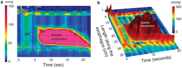
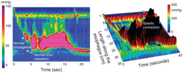
The concept of high-resolution manometry (HRM) is to use sufficient pressure sensors such that intraluminal pressure can be monitored as a continuum along luminal length much as time is viewed as a continuum in conventional manometry. When HRM is coupled with pressure topography plots, pressure amplitude is transformed into spectral colors with isobaric conditions indicated by same-colored regions on the display. Together, these technologies are called high-resolution esophageal pressure topography (HREPT). HREPT has several advantages compared with conventional manometry, the technology that it was designed to replace. (i) The contractility of the entire esophagus can be viewed simultaneously in a uniform format, (ii) standardized objective metrics can be systematically applied for interpretation, and (iii) topographic patterns of contractility are more easily recognized and have greater reproducibility than with conventional manometry. Compared with conventional manometry, HREPT has improved sensitivity for detecting achalasia, largely due to the objectivity and accuracy with which it identifies impaired esophagogastric junction (EGJ) relaxation. In addition, it has led to the subcategorization of achalasia into three clinically relevant subtypes based on the contractile function of the esophageal body: classic achalasia, achalasia with esophageal compression, and spastic achalasia. Headway has also been made in understanding hypercontractile conditions, including diffuse esophageal spasm and a newly described entity, spastic nutcracker. Ultimately, clinical experience will be the judge, but it seems likely that HREPT data, along with its well-defined functional implications, will improve the clinical management of esophageal motility disorders.


Conflict of interest statement

CONFLICT OF INTEREST

Guarantor of the article: Peter J. Kahrilas, MD.

Specific author contributions: Conceived and authored in its entirety.

Potential competing interests: None.

Figures

Figure 1
Figure 1
Typical swallow pressure topography spanning from the pharynx to stomach of a normal subject with normal peristalsis and normal EGJ relaxation imaged as a landscape plot with elevation proportional to the magnitude of intraluminal pressure. As such, the peristaltic contraction appears as a ridge progressing from the upper sphincter to the EGJ over a span of about 10 s. Crucial in the interpretation of pressure topography plots is the assessment of deglutitive EGJ relaxation. This calculation is made within the highlighted rectangular area spanning across the EGJ from the time of the swallow to the arrival of the peristaltic contraction by the method summarized in Figure 2. EGJ, esophagogastric junction.
Figure 2
Figure 2
Detail of the deglutitive EGJ relaxation window illustrating the derivation of the integrated relaxation pressure (IRP). The top panel illustrates a series of pressure profiles across spanning the EGJ at 0.2 s intervals and the blue dot on each indicates the location and magnitude of the greatest pressure along each pressure profile. The lower panel graphs these extracted maximums (blue lines) along with the intermediate values that are not shown in the upper panel. The IRP requires persistence of EGJ relaxation for 4 s within the relaxation window, but the actual time periods that go into its calculation (blue box) can be contiguous, as in this example, or non-contiguous. The IRP was selected as the standard metric for EGJ relaxation because it best differentiated the impaired EGJ relaxation in achalasia from non-achalasic individuals. Adapted from Ghosh et al. (6). EGJ, esophagogastric junction.
Figure 3
Figure 3
Achalasia subtypes are distinguished by three distinct manometric patterns of esophageal body contractility. In classic achalasia (panel a as isobaric contour plot and panel b as a landscape plot), there is no significant pressurization within the body of the esophagus and impaired EGJ relaxation (IRP of 42 mm Hg in this example). The black 42 mm Hg isobaric contour line isolates the portions of the EGJ and UES recording during which the pressure is greater than 42 mm Hg. Panel c represents a swallow from a patient the “achalasia with compression” subtype exhibiting pan-esophageal pressurization. Although high pressures may be recorded in the esophageal body in this subtype, pressure is generated by esophageal shortening in conjunction with contraction of both sphincters, rather than by spasm in the esophageal body. Radiographically, there is no esophageal retention in these patients. Panel d illustrates a pressure topography plot illustrative of spastic achalasia. The three-dimensional rendering of panel d highlights the peaks and valleys of that spastic contraction that would likely appear as a corkscrew pattern on fluoroscopy. These patients often have diffuse thickening of the distal esophageal muscularis propria. EGJ, esophagogastric junction; IRP, integrated relaxation pressure.
Figure 4
Figure 4
Landscape plots of two hiatus hernia patients with functional EGJ obstruction attributable to the crural diaphragm (CD) (a) or the LES (b). In each case, the corresponding IRPCD and IRPLES values are shown. Modified from Scherer et al. (14).
Figure 5
Figure 5
HREPT (a) and landscape plots (b) of an extremely abnormal contraction in a patient with DES illustrating the distinction between rapidly propagated pressurization attributable to intrabolus pressure and to a spastic contraction. The distinction is especially clear in the landscape plot where the individual pressure peaks within the spastic contraction contrast with the straight ridge of intrabolus pressure (labeled IBP) bounded at each end by an area of greater pressure (the UES and crural diaphragm in this instance). This is also the distinction between pan-esophageal pressurization in Figure 3c and distal spasm in three dimensions. Despite the ridge of intrabolus pressure, this swallow has a normal IRP of 11 mm Hg. However, the contractile front velocity of the spastic contraction is very abnormal at 46 cm/s, as is the distal contractile integral (DCI) of the spastic contraction at 36,131 mm Hg•cm• •s.
Figure 6
Figure 6
HREPT (left) and landscape plot (right) of an extremely abnormal contraction in a patient with spastic nutcracker. EGJ relaxation is normal with an IRP of 5 mm Hg. Similarly, the contraction front velocity is normal at 3.2 cm/s. However, the distal contractile integral (DCI), an indicator of the persistence and magnitude of the peristaltic contraction, is more than 10 times normal at 60,300 mm Hg•cm•s. Typical of individuals with these findings, this patient had dysphagia and chest pain.

Similar articles

Cited by

References

    1. Clouse RE, Staiano A. Topography of the esophageal peristaltic pressure wave. Am J Physiol. 1991;261:G677–84. - PubMed
    1. Clouse RE, Staiano A, Alrakawi A, et al. Application of topographical methods to clinical esophageal manometry. Am J Gastroenterol. 2000;95:2720–30. - PubMed
    1. Grubel C, Hiscock R, Hebbard G. Value of spatiotemporal representation of manometric data. Clin Gastroenterol Hepatol. 2008;6:525–30. - PubMed
    1. Spechler SJ, Castell DO. Classification of oesophageal motility abnormalities. Gut. 2001;49:145–51. - PMC - PubMed
    1. Pandolfino JE, Ghosh SK, Zhang Q, et al. Quantifying EGJ morphology and relaxation with high-resolution manometry: a study of 75 asymptomatic volunteers. Am J Physiol. 2006;290:G1033–40. - PubMed